Clinical Experience Tuesday 3rd December 2019 – 4.5 Hours (16:30-21:00): 142 total

Running total of hours: 142

Within this session, I was able to work with returning patients. I enjoy their follow up appointments and their progress interests me a great deal. I feel as though I do not always provide the patient with an effective enough program and do not yet have the confidence in my own ability to correctly prescribe the right volume and intensity of exercises. I understand that much of the time, treatments can be adapted and varied, depending on progress and that trial and error is often an effective means, providing I have adequately and comprehensively reflected on my practice and learnt from any errors. It is not always possible, however to develop an ongoing plan of which can be adapted each session, as the nature of the clinic and the individuals seen do not always allow for follow up appointments. When working with athletes, for example, it is in their best interests to maintain a longer term, continued plan, not only to help reduce pain but to help reduce further risk of injury. With nonathletes and general members of the public however, I have found that often they are satisfied when slight progress is made and as long as their pain subsides and if they are reassured by a diagnosis, then they do not tend to always return.

Patient 1 –
Follow up for insertional achillies tendinopathy
Patient Overview:
Although this patient felt really positive about her previous treatment and noticed considerable change in her symptoms straight afterwards and later on as a result of her exercise prescription, her initial symptoms had returned two days prior to this session and were of similar severity; a sharp pain in her posterior aspect of her heel, most likely the insertion site of the achillies tendon and also around her medial malleolus. Other symptoms, such as the stiffness and feeling of ‘wooden feet’ in the mornings also returned so I felt as though the patient was back at square one with her injury and with no obvious explanation as to why this may have happened, I was left rather confused.
Although the treatment plan on the patients notes on Cliniko suggested a follow up appointment consisting of further sports massage and progression of exercises to facilitate her return to running, because no real progress was made, I decided to conduct another thorough assessment to be sure that my initial diagnosis was still feasible.
From her subjective and objective assessments, it became clear that the tenderness was
more apparent in her tibialis posterior and the patient was adamant that this was exactly the same as her previous treatment, however from the notes and my memory of the session, there was no recollection of this. I did not question the patient and accepted that it will remain unclear as to whether this was a new symptom or not, however it would be reasonable to assume that this irritation of the tibialis posterior may be due to her excellent adherence to her arch strengthening exercises for the treatment of pes planus.
I was unsure as to whether this was a symptom which would be expected or not but was reassured by my supervisor that this pain may start to decrease over time and that we did not have to modify her exercises in the meantime. The tibialis posterior was the cause of the patient’s pain this week and as such it was recommended that STM be performed. When treating this injury in previous appointments, the advice that I was given by my supervisor was to avoid administering any treatment that may irritate the inflamed tendon, instead allow for freer motion and build strength in the associated musculature; addressing the ‘tightness’ in the calf felt by the patient and most likely restricting ankle movements and subsequently creating friction of the structures.
However, on this occasion I had a difference supervisor who advised that I should perform firm deep tissue massage and soft tissue release over the most painful site of pain, with rationale being to desensitise the area to help reduce pain.

I have since tried to find supporting evidence to support this treatment. Although Bowring & Chockalingam (2010) suggested deep tissue massage for tibialis tendinitis, the study also noted that there was no real evidence in support of this for the reduction of pain or increase in function and strength but merely supported theoretical potential for the breaking down of scar tissue and the facilitation of tissue healing. A more recent review by Joseph, Taft, Moskwa and Denegar (2012) which, although some evidence existed on some effectiveness of deep friction massage, the researchers struggled to make a conclusion from their findings. Much of the research was not conducted using deep friction massage as the only treatment modality and so it was suggested that further research was needed in order to test this method alone.
Two years later, Loew et al. (2014) did just that and attempted to test the originally derived theory by Cyriax of deep friction massage treatment for tendinitis, having tested the method on two separate studies consisting of 40 participants with lateral elbow tendinitis and 17 participants with iliotibial band friction syndrome. Of the two studies, neither injuries showed deep friction massage to be effective and although the number of participants in the study was not large, with no significant differences and previous lack of evidence in support of this treatment, I have struggled to find sufficient rationale behind this method.

Conversely, and more specifically, in the case of tibialis posterior tendinosis, a review by Bowring & Chockalingam (2010) recommended rest as a potential treatment for acute tendinopathy as well as the use of orthotics and exercises. At the acute stage, stretching of the gastrocnemius and soleus would aid in increasing dorsiflexion when indicated, however it was suggested that strengthening of the tibialis posterior and should be given when the acute inflammation has subsided.
This could be where this treatment is limiting progression. Because in my objective assessment I found the patient to exhibit pes planus, I wondered whether this may have contributed to the achillies pain, so I prescribed arch strengthening exercises, however this is contraindicated by this review in the early stages of tibialis posterior dysfunction. Whether this pain had only just started to occur from the incorporation of strengthening exercises, or whether it was here from the beginning, it may be advised to stop this exercise for the foreseeable weeks until the inflammation has subsided. The heel raises that I prescribed to help increase eccentric and concentric strength of the achillies tendon and subsequent pain reduction would also activate the tibialis posterior and so this may explain why this caused further irritation to the patient; I will look to reduce these from her exercise routine until the inflammation has subsided also (Bowring & Chockalingam, 2010).

Although strengthening of arch support muscles such as the tibialis posterior and strengthening and stretching of the gastrocnemius and soleus muscles was recommended (Lee & Choi, 2016; Ridge et al., 2018), it may be advisable to wait until the acute swelling and inflammation has subsided and so a period of immobilisation and then controlled mobilisations to increase range of motion may be a more sensible and staged progression in this instance (Bowring & Chockalingam, 2010).

During this treatment, and as advised, I performed two modalities in order to achieve the same outcome. Both of which I tested for effectiveness.
The aim of the treatment was to increase range of motion in the ankle joint, reducing any stiffness and potential friction on the tendons, specifically the achillies and tibialis posterior tendons and surrounding soft tissue. To address the possible joint involvement indicated by the feeling of wooden feet, stiffness and pain in the joints in the mornings, mobilisations with movement was performed to increase dorsiflexion of the ankle joint. There is an abundance of research published, supporting the positive effects on mobilisations with movements for increasing ankle dorsiflexion and reducing pain in individuals with lateral ankle injuries (Loudon, Reiman, Sylvain, 2014; Nisha, Megha and Paresh, 2014) and in knowing that there is also evidence to suggest that limited dorsiflexion can alter running kinematics (Mason-Mackay, Whatman and Reid, 2017), this is a modality worth considering.

To address the possible soft tissue involvement indicated by the feeling of tightness in the patient’s lower posterior legs, soft tissue massage was performed to lengthen the triceps surae muscle group (Stefansson, Brandsson, Langberg and Arnason, 2019).

In order to test which treatment was more effective, we used the knee to wall test, as previously found to be effective as a clinical measure for ankle dorsiflexion and mobility (Hoch & McKeon, 2011; O’Shea & Grafton, 2013).
After the mobilisations with movement, precisely 3 sets of 60seconds, testing in between each set, the patient’s range of movement increased by a third. I then performed the soft tissue release of the tibialis posterior and deep tissue massage of the triceps surae, only to find a reduction in range of motion, most likely due to the irritation of the inflamed tendons and possibly the patient’s apprehension in performing the test due to the increase in pain intensity. This lead to the conclusion that mobilisations with movement was the most effective treatment in this instance and these findings were therefore reflected in her notes, suggesting mobilisations only for a follow up appointment.

However, I am led to question the original rationale behind administering the deep tissue massage and soft tissue release over the painful area, as this was supposed to reduce pain sensitivity in the area but instead, pain levels increased during treatment.
Also, I later noted that after the patient’s previous appointment, improvements in pain and function were reported but that the pain only returned two weeks later, suggesting that the treatment was effective and so It will be of great value to me in my learning to find out whether the more hands on treatment working directly on the inflamed tendon is more effective long term, even if not on during the treatment.
I do understand, however that I have added two variables to the rehabilitation, deep tissue massage directly over the tibialis posterior and insertion area of achillies, as well as mobilisations. If the patient reports improvements in her next session, I will be unlikely to be able to differentiate between the two treatments.

Patient 2 – Unfortunately, my second patient did not arrive and this was the second session that he missed. I was made aware of the traffic delays in the city, so hope that his was a factor but had hoped that basic politeness would result in a courtesy call of apology.
I have noticed that over the past month, the clinic has experienced a number of cancellations at the last minute, giving little or no chance for the therapists to arrange for alternative appointments. It is within the clinic policy for the patient to provide notification of a cancellation at least six hours in advance. I am sympathetic to the fact that there are some occasions that are unavoidable and that sometimes any notification is impossible or that sometimes mistakes happen and that appointments get forgotten. In these occasions, businesses have to accept the loss of income or wasted time. However as this seems to be becoming a common occurrence, it may be useful to make alterations to the policy. It has been suggested that although there is no set time of which patients should notify cancelations, if no-shows are common then any increases in policy time could serve to reduce those instances (Huang & Zuniga, 2014).
As a group of five students without patients in this hour of the session, we all gathered around together to debate and discuss efficacy of soft tissue massage and it’s role within the clinic environment or sports therapy and rehabilitation. I have reasonably strong opinions on the psychosocial benefits of massage in any form, as often made obvious within this blog, regardless of the physiological aspects of this treatment and was able to provide some references for this, which will serve me well when discussing this area of treatment with patients in the future.
I found this type of ‘debate’ environment very useful, as it mimicked how I would imagine a discussion would develop on this type of subject with a patient or another health care professional, who perhaps needs extra understanding on our role as sports therapists or on our treatment rationale.

Free hour between patients – I used this time to catch up on my notes from my previous session and was able to join another student sports therapist who was conducting an initial appointment for shoulder pathology. This is an area that I find challenging, so I was happy to work together with another student to help diagnose this patient with possible rotator cuff tendinopathy and offer my skills in developing an effective strengthening program, which is something that I do feel confident doing.

Patient 3 – 13 year old rugby player, pain in thoracic spine, specifically on palpating T2.
This was another patient under the age of 16 who attended the clinic with his parent. I felt a little more comfortable treating this patient, having experienced treating a child the day before, however, fortunately on this occasion, the parent sat in waiting room and left the patient to be assessed by himself. I felt much more natural speaking with the patient on a one-on-one environment as opposed to balancing the conversation between myself, parent and child and felt as though the patient could open up more to me without the judgement or interruptions from his parent.
The patient was experiencing upper thoracic pain and on palpation, we established that it was on the spinous process of T2 specifically. This pain was noticed since a heavy weekend of playing and now occurs during physical activity and sometimes at rest. After seeing his team physiotherapist, who the patient reports as too busy to see for the foreseeable, he was assured that it was tight muscles and was advised to visit a sports therapist for a soft tissue massage.
However, although I wanted to oblige to his request of a soft tissue massage, I wanted to be sure that there was no underlying issue that could be contributing to his pain rather than just treating the symptoms. I conducted an assessment, with the patient’s permission. Initially, I did not find a reason for the pain and was confused as to why the most painful palpable area was on the spinous process.
Baffled, I requested the assistance of the clinic supervisor who, on entering the cubicle immediately spotted his hyper lordotic seated posture, of which was the most pronounced that I had ever seen. I was very surprised that this had not occurred to me and that I had not noticed this, especially as I had recently attended an informative lecture on posture and shoulder/thoracic pain. I can only think that the reason this was missed, is because the patient was so young and that not only my observational skills and common sense were clearly lacking in this assessment, but that I had unconscious predisposed misconceptions that poor posture is developed over time and not present in children so young. Now that I know the possible benefits of considering alterations in posture when addressing shoulder and thoracic pain, I would be doing my patients huge injustices if I made this incorrect assumption regarding the younger individuals; correcting poor posture early on could prove paramount in future risk factors for injury, serving to prevent muscle imbalances, skeletal deformities and developmental dysfunctions, with early poor posture habits contributing to future deterioration and musckoloskeletal strain (Kim, Cho, Park and Yang, 2015).

One of the most useful exercises I learnt from the aforementioned shoulder lecture was the method of asking a patient to place their finger on their sternum then using their chest to push those fingers away as a means to inadvertently correct their shoulder posture. I found this incredibly useful in helping my young patient acquire a method to remind himself of the posture that he would be aiming to achieve through re-education and strengthening; just one motion created the desired effect and with the need to adjust technical language for younger patients to understand, this was a very useful tool.

I was also able to use other information acquired from the shoulder lecture regarding hyper kyphosis as this was the first part of the shoulder symptom modification procedure (Lewis, 2009; Lewis, 2011). I found this video by the Physio tutors very helpful in understanding this procedure: https://www.youtube.com/watch?v=sYTW7u6ZoCI.
As modifying this patient’s thoracic spine by way of reducing the kyphotic curve, reduced his symptoms, according to this procedure, this indicated the need to focus on strengthening the thoracic spine and as such, I looked to prescribe some of these newly learnt techniques to help reduce this. I prescribed a posterior capsule stretch, as recommended by Lewis (2009) and attempted to recommend strengthening. Although I have a wide range of mobility exercises for the thoracic spine of which I was able to show the patient, including the lawn mower and threading the needle, I only had one exercise on building muscular strength for the back muscles; the low row specifically targeting the rhomboids and lower and middle traps. It is especially important that I develop and repertoire of exercises, especially ones that require no specialist equipment, for patients who do not have access to a gym to use a row machine.

I found an excellent video demonstrating a good exercise that can be done at home and one I feel more comfortable prescribing in the future.

References –

Bowring, B., & Chockalingam, N. (2010). Conservative treatment of tibialis posterior tendon dysfunction-A review. Foot. https://doi.org/10.1016/j.foot.2009.11.001

Hoch, M. C., & McKeon, P. O. (2011). Normative range of weight-bearing lunge test performance asymmetry in healthy adults. Manual Therapy, 16(5), 516–519. https://doi.org/10.1016/j.math.2011.02.012

Huang, Y. L., & Zuniga, P. (2014). Effective cancellation policy to reduce the negative impact of patient no-show. Journal of the Operational Research Society. https://doi.org/10.1057/jors.2013.1

Joseph, M. F., Taft, K., Moskwa, M., & Denegar, C. R. (2012). Deep friction massage to treat tendinopathy: A systematic review of a classic treatment in the face of a new paradigm of understanding. Journal of Sport Rehabilitation. https://doi.org/10.1123/jsr.21.4.343

Kim, D., Cho, M., Park, Y., & Yang, Y. (2015). Effect of an exercise program for posture correction on musculoskeletal pain. Journal of Physical Therapy Science, 27(6), 1791–1794. https://doi.org/10.1589/jpts.27.1791

Lee, D., & Choi, J. (2016). The Effects of Foot Intrinsic Muscle and Tibialis Posterior Strengthening Exercise on Plantar Pressure and Dynamic Balance in Adults Flexible Pes Planus. Physical Therapy Korea, 23(4), 27–37. https://doi.org/10.12674/ptk.2016.23.4.027

Lewis, J. S. (2009). Rotator cuff tendinopathy/subacromial impingement syndrome: Is it time for a new method of assessment? British Journal of Sports Medicine, 43(4), 259–264. https://doi.org/10.1136/bjsm.2008.052183

Lewis, J. S. (2011). Shoulder Symptom Modification Procedure ( SSMP ) V2 Date : Symptomatic movement or posture 1 : Symptomatic movement or posture 2 : (1), 2011.

Loew, L. M., Brosseau, L., Tugwell, P., Wells, G. A., Welch, V., Shea, B., … Rahman, P. (2014). Deep transverse friction massage for treating lateral elbow or lateral knee tendinitis. Cochrane Database of Systematic Reviews. https://doi.org/10.1002/14651858.CD003528.pub2

Loudon, J. K., Reiman, M. P., & Sylvain, J. (2014). The efficacy of manual joint mobilisation/manipulation in treatment of lateral ankle sprains: A systematic review. British Journal of Sports Medicine, 48(5), 365–370. https://doi.org/10.1136/bjsports-2013-092763

Mason-Mackay, A. R., Whatman, C., & Reid, D. (2017). The effect of reduced ankle dorsiflexion on lower extremity mechanics during landing: A systematic review. Journal of Science and Medicine in Sport, 20(5), 451–458. https://doi.org/10.1016/j.jsams.2015.06.006

Nisha, K., Megha, N. A., & Paresh, P. (2014) Efficacy of weight bearing distal tibiofibular joint mobilization with movement (MWM) in improving pain, dorsiflexion range and function in patients with post acute lateral ankle sprain quick response code. Int j physiother res.

O’Shea, S., & Grafton, K. (2013). The intra and inter-rater reliability of a modified weight-bearing lunge measure of ankle dorsiflexion. Manual Therapy. https://doi.org/10.1016/j.math.2012.08.007

Ridge, S., Henderson, A., Bruening, D., Jurgensmeier, K., Olsen, M., Griffin, D., … Davis, I. (2018). Midfoot Angle Changes During Running After an 8-week Foot Strengthening Program. Foot & Ankle Orthopaedics, 3(3), 2473011418S0040. https://doi.org/10.1177/2473011418s00405

Stefansson, S. H., Brandsson, S., Langberg, H., & Arnason, A. (2019). Using Pressure Massage for Achilles Tendinopathy: A Single-Blind, Randomized Controlled Trial Comparing a Novel Treatment Versus an Eccentric Exercise Protocol. Orthopaedic Journal of Sports Medicine, 7(3), 1–10. https://doi.org/10.1177/2325967119834284


Clinical Experience Monday 2nd December 2019 – 5 hours (15:00-20:00): 137.5 total

Running total of hours: 137.5

Unfortunately, this session was somewhat setback by an ongoing lower back issue of mine and so I was not able to fully apply myself physically to administer any manual treatments, however as I really enjoy attending the clinic and feel a responsibility for my patients I did not want to miss the opportunity to treat and attended to gain further practical experience, but with occasional manual help when needed; to start with I worked with another student for 2 patients for the first part of this session until I felt I could manage, offering advice where I could and taking an equal part in the appointments.

Patient 1 – Neck Injury and STM
This patient was a student who rugby player sustained a neck injury during a game one week ago and this was her follow up appointment.
It is reported that concussion, most likely occurring during a tackle, is the most common injury in rugby union (Cross et al., 2019).
Initially I was anxious about assessing a neck injury but by going through her previous notes, I was reassured by her previous appointment with Alex Walker, the clinic supervisor; the notes were thorough and clearly stated the need to revisit A & E, where the patient was further assessed and any serious injury was ruled out.
The first assessment was performed one day after the injury and it was obvious that the patient was concussed. Because of this and coupled with the very acute nature of her symptoms, no tests were performed, however her visit to A & E ruled out any neurological involvement and a thorough test was carried out.
If this was my patient alone, I would probably have subjected her to a full assessment, including any neurodynamic testing of the cervical spine, however the therapist in this appointment chose not to do this based on the absence of any indicative symptoms. I would agree that this is good rationale for not putting this patient through another session of testing, as she had already been subjected to a number of assessments within the week prior to this session and has simply requested a STM.
The muscle testing for this patient was strongly indicative of muscular tension and resultant pain, so this coupled with the mechanism of injury and with the improving nature of the pain and ROM, a deep tissue massage with passive and active passive stretching was performed to help facilitate the increase in ROM.
I often forget to include objective measures within my treatment, but in this instance, because I perform ROM tests as part of the basic assessment, I am able to use this as a marker, as long as I remember to retest at the end of the treatment.
This patient reported increases in pain and ROM after the massage and stretching treatment and as such, we knew that this treatment was the most effective treatment for her and prescribed some further stretches to complete at home, as well as a strengthening program for her to start when ROM is back to full range and when pain has mostly subsided.
We felt confident that this individual would adhere to any home exercise programs prescribed because of her passion to return to rugby and her need to be fit to play again.
We also discussed the importance of taking the advised time off of her sport, due to her concussion. It is important to educate individuals about the dangers and risk factors involved with concussion and to be aware of a number of factors associated with this type of traumatic injury. A study by Bussey et al. (2019) found that neuromuscular control can be altered during rugby tackling within twelve months of sustaining a concussion which can pose as a potential future risk factor for subsequent concussions.
Initially, I had naively thought that by increasing muscular strength of the neck muscles, future injury or concussion risk may be reduced, however a review by Benson et al. (2013) found no evidence in support of this, nor any evidence advocating the use of mouth guards or helmets. A recent study suggested that tackling position and acceleration and speed of the tackler can be altered as a way to reduce the risk (Cross et al., 2019) but these as external factors that are beyond the control of the therapist. From this, the only advice that I was able to offer this patient was to follow guidelines provided by the national governing body of her sport, in this case rugby and to take the suggested time off by the physicians at the hospital.

Patient 2 – STM and Traction at hip
This patient was returning for another session of STM of the back with additional piriformis release and manual hip traction.
After his session three weeks previous, he remarked on his improvements, having felt a significant relief from his usual tension.
After my previous session with this patient on 11th November, I looked into the value of this treatment and ways in which this could be done at home to save his time and money. Reiman and Matheson (2013) supported the use of a home-made device that simply connects the foot with an anchor, such as heavy furniture or gym equipment so that the individuals can apply a long axis distraction force of the hip. I suggested this option to my patient so that he could start to rely less on treatments and self-manage his discomfort, however he immediately responded with his dissatisfaction at any self-performed treatment and explained that he was aware that he could potentially administer his own treatment.
It is in my opinion that as long as the patient is aware of the transient nature of the STM and traction and as long as we, as therapists, provide the patient with exercises to progress through and educate them on their injury and rehabilitation plan, if they continue to want these passive treatments then this is completely justified. It would be unethical, however if I were to provide this basic level of treatment on its own, unaccompanied by a more long term correction and patient education.
I am aware of the short term, transient nature of soft tissue treatment and the need to incorporate strengthening exercises as the most part of any rehabilitation plan for developing and enhancing functional movements, however I am also of the opinion that these two are transient; when exercises are stopped, muscles become weakened again. So, in my view both strengthening and passive soft tissue therapy are transient, the difference is that the patient is more able to perform the strengthening exercises at home and become fully empowered to sustain their own musculoskeletal function, compared with the more specific requirements of STM and the need to see a specialist for a more satisfying experience, as experienced by this patient.
People pay to get their hair or nails treated, but there are no long term benefits of this to their health, so as long as people are attending their appointments knowing that they are receiving treatment to help with their pain and ROM in the short term then I feel like the treatment is entirely justified. We should always provide the patients with information about how they might be able to maintain their function outside of the clinic but also we must be aware that this may be of no interest to some patients and that short term relief is their only need.

Patient 3 – Osgood’s Schlatter’s Disease
This was my first experience of treating a child under 16 and although initially I felt uncomfortable, I soon became more confident and was able to communicate well with both the patient and his mother.
I was aware that I needed to keep my language simple and any complicated jargon to a minimum. I did find myself talking to the patient’s mother on more occasions that I think was necessary however and as such the patient may have felt uninvolved in the assessment procedure. I was able to pick up on this early enough and changed my approach, which soon brought the attention back to the young patient, who may then have felt more empowered in this initial stage in his rehabilitation.
It became apparent early on, just from the subjective assessment that Osgood’s Schlatter’s disease (OSD) was the most likely diagnosis; bony prominence in both tibial tuberosities, pain in all dynamic sporting movements, high volume of exercise, specifically football, pain when kneeling and the age of the patient being 13 (with the most likely age in the develop of this condition being between 10 and 15 (Vaishya, Azizi, Agarwal and Vijay, 2016)). I continued to conduct a throughout assessment, remembering to clear joints above and below and then felt the need to seek additional advice from a supervisor, as this patient was so young.
When the supervisor initially joined us in the assessment process to offer further advice, the first thing mentioned was the bony ‘lumps’ under both of the patient’s knees. It was quite abruptly mentioned that the lumps will stay but the pain will go, to which both the mother and the patient were taken aback. At first I was surprised at their reactions but now understand why this may have caused some distress; the lumps in the patients’ knees are quite obvious and if they had always visualised this to reduce over time, to find out that the lumps would remain, would have come as a shock. I did take the time later in the appointment to explain that some of the prominence may have been down to acute inflammation, effusion and heat and also more obvious due to the lack of muscle tone in the patient’s leg and from this conversation, both the patient and his mother felt more relaxed about this aesthetic element to the injury. In cases where those prominences are extreme, there is a procedure known as closing-wedge osteotomy, which was found to be an effective means of managing the bony prominence on the tibial tuberosity which is formed as a result of Osgood-Schlatter’s disease (Pagenstert, Wurm, Gehmert and Egloff, 2017).

Figure 1 shows the location and aesthetic appearance of the bony prominence of Osgood’s Schlatter Disease, as well as an x-ray image (Vaishya et al., 2016 p.4;p.5).

Interestingly, only 25% of individuals report pain in the tibial tuberosity (Vaishya et al., 2016) so this is not necessarily the only symptom to consider in this condition.

Osgood’s Schlatter’s Disease is characterised by the inflammation of the patella tendon, precisely over it’s insertion into the tibial tuberosity (Vaishya et al., 2016).
Pathogenic risk factors for OSD are vast, with Watanabe et al. (2018) reporting the following all possible contributors to the disease;
1. Height
2. Weight
3. BMI
4. Quadriceps tightness in kicking leg
5. Soleus, gastrocnemius and quadriceps tightness in non kicking, support leg
6. Centre of gravity
7. Medial Longitudinal Arch measurement

It has been found in a study on 20 subjects with a mean age of 13.4 years old, the same age as the patient that stretching of the quadriceps muscles, in particular, the rectus femoris would help to reduce OSD symptoms (Tzalach et al., 2016), however when I performed the Thomas Test it showed nothing but excellent flexibility in the patient’s hip flexors and knee extensors, specifically the quadriceps.

I have since read, however that although the modified Thomas test, shown in figure 1 has been found to be a reliable measure of hip flexor flexibility, it is reliant of a controlled pelvic tilt (Vigotsky et al., 2016).
I do not recall having taken pelvic tilt into account on this occasion, so may not be able to use this as a valid test in this instance unless I retested to account for this variable. However, just from observing, I would be comfortable in suggesting that quadricep flexibility was probably not an issue for this individual.
Instead, we prescribed exercises to strengthen gluteal muscles and hamstrings and educated him and his mother on workload and advised to incorporate plenty of non-sporting days to facilitate his growth but without applying excessive loads. Vaishya et al. (2016) also recommended quadriceps strengthening in addition to the above, so if this patient were to return, I would look to incorporate this element into the rehabilitation program, considering also the lengthened muscles in this case; this may be an indicator of weakness, as opposed to flexibility and I should have taken this into account when assessing this.

With the aim to reduce pain and manage swelling, we advised him to treat any inflammation as and when needed and to continue in his use of ICE and another other relieving modalities, which may also help to reduce the inflammation of the OSD but also of other conditions that may also be present or likely in this particular case, such as patella tendinitis or infrapatellar bursitis, as reported by (Vaishya et al., 2016).
Often, the presence of both of these aforementioned conditions are likely alongside OSD but sometimes these conditions may be the cause of the symptoms but mistaken for OSD; of these, however, it is hard to make an accurate differential diagnosis due to their similar epidemiology and presentation although treatment is often similar (Vaishya et al., 2016). In this case, however, the obvious prominence, an example shown in figure 1, allows for us to assume OSD as the primary condition but be aware of and treat any secondary issues that may arise.

It was also advised that surgery may be viable option, should conservative treatment not be effective and if the condition remains restrictive and debilitating (Vaishya et al., 2016), however this is rare and usually the symptoms reduce or diminish over time and as previously mentioned, only the prominence will remain.

It will be interesting to know whether the symptoms do start to resolve over the coming weeks, in spite of his plan to continue in his activity levels; he seemed (and understandably so) reluctant to reduce his football hours and so progress may be slower. However, with the exercises prescribed and after receiving information on the condition, the patient and his mother may have a better awareness of how they are able to manage the condition, should the symptoms worsen.

Extra Research in my final hour in clinic – 
Initially, after his mother informed us that the patient had been taking Non-steroidal Antiinflammatory Drugs (NSAIDS), specifically Advil, in order to continue to play, I was concerned and was unsure as to whether this was the most appropriate course of treatment to take. As I had no prior knowledge on this, I did not provide any further advice to either advocate the use of these or otherwise. However, according to recent research on OSD, NSAIDS are suggested (Kolodychuk, 2018) and as such perhaps a personal choice by parents of which is beyond my scope of knowledge or area to advice.

References –

Benson, B. W., McIntosh, A. S., Maddocks, D., Herring, S. A., Raftery, M., & Dvořák, J. (2013). What are the most effective risk-reduction strategies in sport concussion? British Journal of Sports Medicine. https://doi.org/10.1136/bjsports-2013-092216

Bussey, M. D., McLean, M., Pinfold, J., Anderson, N., Kiely, R., Romanchuk, J., & Salmon, D. (2019). History of concussion is associated with higher head acceleration and reduced cervical muscle activity during simulated rugby tackle: An exploratory study. Physical Therapy in Sport. https://doi.org/10.1016/j.ptsp.2019.03.012

Cross, M. J., Tucker, R., Raftery, M., Hester, B., Williams, S., Stokes, K. A., … Kemp, S. (2019). Tackling concussion in professional rugby union: A case-control study of tackle-based risk factors and recommendations for primary prevention. British Journal of Sports Medicine. https://doi.org/10.1136/bjsports-2017-097912

Kolodychuk, N. (2018). Tendinosis as the under lying pathology of osgood-schlatter disease: imaging similarities and treatment implications. International Journal of Orthopaedics Sciences. https://doi.org/10.22271/ortho.2018.v4.i1j.97

Pagenstert, G., Wurm, M., Gehmert, S., & Egloff, C. (2017). Reduction Osteotomy of the Prominent Tibial Tubercle After Osgood-Schlatter Disease. Arthroscopy – Journal of Arthroscopic and Related Surgery. https://doi.org/10.1016/j.arthro.2017.02.012

Reiman, M. P., & Matheson, J. W. (2013). Restricted hip mobility: clinical suggestions for self-mobilization and muscle re-education. International Journal of Sports Physical Therapy.

Tzalach, A., Lifshitz, L., Yaniv, M., Kurz, I., & Kalichman, L. (2016). The Correlation between Knee Flexion Lower Range of Motion and Osgood-Schlatter’s Syndrome among Adolescent Soccer Players. British Journal of Medicine and Medical Research. https://doi.org/10.9734/bjmmr/2016/20753

Vaishya, R., Azizi, A. T., Agarwal, A. K., & Vijay, V. (2016). Apophysitis of the Tibial Tuberosity (Osgood-Schlatter Disease): A Review. Cureus, 8(9). https://doi.org/10.7759/cureus.780

Vigotsky, A. D., Lehman, G. J., Beardsley, C., Contreras, B., Chung, B., & Feser, E. H. (2016). The modified Thomas test is not a valid measure of hip extension unless pelvic tilt is controlled. PeerJ. https://doi.org/10.7717/peerj.2325

Watanabe, H., Fujii, M., Yoshimoto, M., Abe, H., Toda, N., Higashiyama, R., & Takahira, N. (2018). Pathogenic Factors Associated With Osgood-Schlatter Disease in Adolescent Male Soccer Players: A Prospective Cohort Study. Orthopaedic Journal of Sports Medicine, 6(8), 1–8. https://doi.org/10.1177/2325967118792192

External Placement Exmoor Osteopaths Thursday 28th November 2019 – 4 hours (09:00-13:00): 132.5 total

Running total of hours: 132.5

During my session at the Osteopath clinic, Kat and I spent the majority of time with returning patients requiring follow up treatments, all of which self-reporting improvements. Due to the demographics in the area of the clinic in Minehead, most patients attending the clinic are of the older generation and from conversing with them and from hearing Kat’s experiences, they seem more reluctant to engage in any exercise program away from the clinic and although they accept this as a huge limiting factor in their progress, they also seem to be under the false understanding that as they are of age, they cannot improve their physical status and are happy to visit the clinic on a regular basis for passive treatment.
As much as I am an advocate for the use of massage, even if the benefits are psychological due to the absence of a stock of high quality evidence, I truly believe that their quality of life will significantly improve if they became more active in their own treatment.
I wonder if because Kat has now been treating the same individuals over a relatively long length of time, having been at the clinic for over seven years, her attempts at encouraging some of the individuals to be more proactive have lessened.
In my final three weeks here at this clinic, I hope to instil a sense of belief into those particular clients, reinforcing the benefits that may arise should they start to incorporate strengthening, even if only generalised and nonspecific, into their everyday lives.
I could attempt to speak with the clients about why they feel they cannot become more physical active and look to come up with strategies to encourage exercise adherence, such as finding ways to incorporate exercises into their daily routines, as found to be effective by Bassett (2015).
With one particular patient during this session, gardening was their means of physical activity, but was also one of the main causes for the onset of pain. With patient education and endorsement of better technique and adding in extra movements, such as side walking, squatting for gluteus muscles, the conveniently named lawn mower or chopping manoeuvres for thoracic strengthening, then the everyday activity becomes more specific but yet without any perceived additional effort.

I will also look to provide each patient with a new specific exercise plan after each treatment and discuss a way individualised to them, that may enhance their cooperation.
I do not want to undermine the current treatment plan from Kat and must ensure that the exercises I prescribe are suitable and positively facilitate this, so I will be sure to consult with both the patient and Kat beforehand.

Of all the patients we saw during this morning session, including sacroiliac joint pain and dysfunction, non-specific lower back pain and shoulder tendinopathy (although now I may have approached this differently, having read more into shoulder kinematics and scapular dyskinesis), the highlight of this session for me, was discovering a new injury of the spine, Spondylolisthesis, of which I reluctantly admit, have never heard of before.
I was unsure as to the exact pathology in this patient and as such required additional assistance from the Osteopath. Very early on, the word “Spondy” was thrown around as though it were a common injury and as such I did not show my lack of knowledge in front of the patient by asking for clarity, instead I waited until the end of the consultation to question Kat. In hindsight, I should have expressed my willingness to learn something knew and rise above my lack of knowledge as it would have been really informative to have gone through the basics, for example just palpating the vertebrae; in future there will be instances when I will not know the issue and in these cases, so the more open and honest I am with the client, the more trusting they will be of my integrity and ability to provide an honest and evidence based treatment.
I took the time outside of the clinic to research Spondylolysis and how I may be able to assess for this in the future.
I needed to identify the symptoms that would indicate this and how I would be able to treat the patient.
It is advised that in cases of radiculopathy whether back pain is present or not, spondylolysis should be considered (Watters et al., 2009). Due to the age of this patient, it is most likely that this is of a degenerative nature and so in this instance, pain may be minimal or less apparent.
According to Watters et al. (2009. p.611) spondylolysis is the “anterior displacement of one vertebra over the subjacent vertebra, associated with degenerative changes, without an associated disruption or defect in the vertebral ring”.
In this patient, the displacement occurred at L2 and I was able to briefly palpate a protrusion. At first I thought this protrusion could be of the affected vertebrae, but in fact this vertebrae only feels protruded relative to the anteriorly displaced one above. The most accurate way to diagnose this condition is radiography and as reported by Watters et al. (2009), this condition often presents itself alongside (and is often thought to cause) spinal stenosis (Zhu et al., 2017), which can be further identified by magnetic resonance imaging (MRI) with surgical treatment by way of decompression being the most supported option among specialists. In a study by Weinstein, Dolan, Wright and Dobbs (2013), in which the non-operative treatment was compared with the conservative treatment for this condition, improvements in pain and function were greater experienced in the operative group for up to four years post surgery.
Although GP referral would be a considered as a likely course of action for myself as a therapist, recent research by Zhu et al. (2017) recommends application of conservative treatment first but also highlights the lack of understanding in the mechanisms behind the development of this condition and as such a specific focus on treatment has not been fully developed. Zhu et al. Strengthening and stretching exercises of the extensors and flexors of the spine to develop and maintain spinal stability are advised by Zhu et al. (2017), which addresses the lack of stability in the spine which has been commonly reported as a contributing factor.
It is also suggested that lower discogenic back pain could result in weakened spinal stabilising muscles and therefore eventually lead to degenerative spondylolysis (Zhu et al., 2017), allowing the assumption that strengthening these structures could improve the overall stability of the spine and associated pain.

Having had the opportunity to experience a clinical suspected diagnosis of spondylolysis and with my better understanding behind the mechanical presentation of the condition, I would be more aware of the need to consider this within future assessments, especially in the older population.

References –

Bassett, S. (2015). Bridging the intention-behaviour gap with behaviour change strategies for physiotherapy rehabilitation non-adherence. New Zealand Journal of Physiotherapy, 43(3), 105–111. https://doi.org/10.15619/nzjp/43.3.05

Watters, W. C., Bono, C. M., Gilbert, T. J., Kreiner, D. S., Mazanec, D. J., Shaffer, W. O., … Toton, J. F. (2009). An evidence-based clinical guideline for the diagnosis and treatment of degenerative lumbar spondylolisthesis†. Spine Journal, 9(7), 609–614. https://doi.org/10.1016/j.spinee.2009.03.016

Weinstein, S. L., Dolan, L. A., Wright, J. G., & Dobbs, M. B. (2013). Effects of bracing in adolescents with idiopathic scoliosis. New England Journal of Medicine. https://doi.org/10.1056/NEJMoa1307337

Zhu, R., Niu, W. xin, Zeng, Z. li, Tong, J. hua, Zhen, Z. wei, Zhou, S., … Cheng, L. ming. (2017). The effects of muscle weakness on degenerative spondylolisthesis: A finite element study. Clinical Biomechanics, 41, 34–38. https://doi.org/10.1016/j.clinbiomech.2016.11.007

Clinical Experience Tuesday 26th November 2019 – 2 hours (15:00-20:00): 128.5 total

Running total of hours: 128.5

Patient 1 – For my first patient of the session I performed a soft tissue massage treatment for a frequent visitor to the clinic, with this being her sixth session. The assessment was very similar to previous sessions in that small ROM differences were observed and treatment options were discussed; I wanted to ensure that the patient was aware of the transient nature of the massage and that there are other options to be considered, such as mobilisations and exercises. However, the patient was happy to receive a STM only, as this was all she was comfortable with. From previous sessions, I have become aware of the psychosocial element to this patient’s clinic visits and as such feel more than happy to accommodate her in this; by coming in on a weekly basis, this elderly woman has a means to talk about her week and to offload her difficult current situation in a confidential environment in which I am able to provide her with my under divided attention and genuine regard for her wellbeing and happiness. I feel like I have developed a positive and friendly relationship with my patient and I hope I am enhancing her week by being able to listen to her with care. The patient has tried to complete her rehabilitation exercises at home and small improvements to her ROM have been noticed, however not as much as I would like. I would hope to facilitate changes in her physical, as well as her mental wellbeing but I also know that this may take time and perhaps a change in one may lead to a change in the other.
I will continue to encourage her participation in her prescribed home based exercises and advocate their benefits in the hope that she will have a better quality of life through better ROM of her shoulders and neck.

Patient 2 – During my clinical experience in my previous session, I applied kinesiology tape, now known as elastic Therapeutic Taping (ETT) to the patient’s shoulder as requested and as advised by the patient’s previous therapist. The patient reported the tape as beneficial by way of reminding him to correct his posture (mechanoreceptors) and he felt his pain was subsequently reduced.
I applied the tape with full stretch from anterior to posterior without much regard for the location; the patient was in full stretch of his pectoralis muscle group with shoulders as far back as possible, so the tape, regardless of location, would serve to act as postural taping.
I am not confident in my opinion or understanding as to the effectiveness of ETT and therefore efficacy of its and therefore I took the time to conduct some research on this topic, in order to be able to advocate its use in clinic, but also so that I can educate the patient more confidently about the rationale behind this treatment.

Although there are some supported claims of the benefits of kinesiology tape, such as increase in strength, range of motion and proprioception, there is not enough high quality evidence to support its widespread popularity and use in the sports injury context (Williams et al., 2012).
In the treatment of musculoskeletal injuries, a systematic review concluded that ETT did not have enough evidence in support of its use on sports injuries, but that potential perceived effects were noted (Mostafavifar et al., 2013).
Konishi (2013) found that ETT acted to compensate muscle weakness, proposedly by affecting afferent neurophysiology by way of sensory input. This study consisted of two groups, a taping group and a non-taping group with the results showing greater muscle contraction in the taped muscle compared with the non-taped muscles of the knee, particularly the quadriceps femoris. Although these findings seem to support the use of ETT in injuries presenting with muscle weakness, in this particular case of the knee, the design of the study, whereby the control group were not taped, allowed for the potential of the tape having a placebo affect on the intervention group, i.e. the presence of tape alone was enough to benefit the subject.
The suggestion of ETT having a placebo effect is long debated and the subject of controversy and conflicting research. A study by Poon et al. (2015) claimed that any positive effects of ETT should be assumed the result of a placebo effect, with this study finding that this tape did not enhance or assist in muscle strength and performance of the quadriceps.
However, a single-blinded randomized control pilot study in the same year by Han et al. (2015) was conducted on 14 male office workers with rounded shoulder posture using a control group, providing some evidence of its effects by ruling out possible placebo and crediting any positive benefits on forward shoulder posture and increased pectoralis minor length to its mechanical support; the control group subjects had tape applied but with no stretch whereas the intervention group had 35-40% stretch.
More recent research by Shih et al. (2017) also found that both exercises and ETT provided positive improvements to forward head posture, with no significant differences between the two treatments, as conducted on a study on 60 subjects.

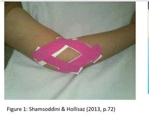
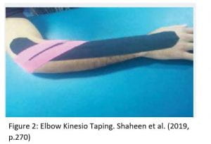
Although the purpose or benefits of ETT are unclear and although there is not enough supporting evidence on this treatment in the management of sports injuries or enhancing sports performance, there are studies providing high quality evidence supporting its use on specific sports injuries, such as lateral epicondylitis. In particular, a study by Shamsoddini & Hollisaz (2013) reported a significant difference in grip strength and extension force as well as the reduction in pain in 30 subjects with lateral epicondylitis, compared to the same treatment on the unaffected arms, which provided control data). Another study on the effects of both ETT and therapeutic ultrasound on lateral epicondylitis, finding that this taping modality did have significant effects on pain and more so than the ultrasound (Shaheen et al., 2019).

More specifically, in the treatment of shoulder injuries, the only research I found supporting its effectiveness in postural correction were case studies and provided very poor evidence for its use. However, from the research I have found, as ETT is not likely to cause any harm to the patient, I would consider using this as a treatment method for any control of pain or posture, where neurology becomes a factor and continue to use it on a case by case basis; if the patient reports improvements in their symptoms or performance, then I would continue to use it in their treatment program.

References –

Han, J. T., Lee, J. H., & Yoon, C. H. (2015). The mechanical effect of kinesiology tape on rounded shoulder posture in seated male workers: A single-blinded randomized controlled pilot study. Physiotherapy Theory and Practice. https://doi.org/10.3109/09593985.2014.960054

Konishi, Y. (2013). Tactile stimulation with Kinesiology tape alleviates muscle weakness attributable to attenuation of Ia afferents. Journal of Science and Medicine in Sport. https://doi.org/10.1016/j.jsams.2012.04.007

Mostafavifar, M., Wertz, J., & Borchers, J. (2013). A systematic review of the effectiveness of kinesio taping for musculoskeletal injury. Physician and Sportsmedicine. https://doi.org/10.3810/psm.2012.11.1986

Poon, K. Y., Li, S. M., Roper, M. G., Wong, M. K. M., Wong, O., & Cheung, R. T. H. (2015). Kinesiology tape does not facilitate muscle performance: A deceptive controlled trial. Manual Therapy. https://doi.org/10.1016/j.math.2014.07.013

Shaheen, H., Alarab, A., & S Ahmad, M. (2019). Effectiveness of therapeutic ultrasound and kinesio tape in treatment of tennis elbow. Journal of Novel Physiotherapy and Rehabilitation, 3(1), 025–033. https://doi.org/10.29328/journal.jnpr.1001025

Shamsoddini, A., & Hollisaz, M. T. (2013). Effects of taping on pain, grip strength and wrist extension force in patients with tennis elbow. Trauma Monthly. https://doi.org/10.5812/traumamon.12450

Shih, H. S., Chen, S. S., Cheng, S. C., Chang, H. W., Wu, P. R., Yang, J. S., … Tsou, J. Y. (2017). Effects of Kinesio taping and exercise on forward head posture. Journal of Back and Musculoskeletal Rehabilitation. https://doi.org/10.3233/BMR-150346

Williams, S., Whatman, C., Hume, P. A., & Sheerin, K. (2012). Kinesio Taping in Treatment and Prevention of Sports Injuries. Sports Medicine. https://doi.org/10.2165/11594960-000000000-00000

Clinical Exprience Monday 25th November 2019 – 5 Hours (15:30-20:00): 126.5 total

Running total of hours: 126.5

Patient 1 – The first patient in this session did not attend his appointment, so in order to fully utilise my time within the clinic, I shadowed a post graduate student; this was a fantastic opportunity for me to offer any of the knowledge that I have developed from the previous week of shoulder treatments and to assist where possible.
This patient experienced sharp non localised pain in his shoulder joint when bringing his arm up above his shoulder passed 90degrees of flexion and in active internal and external rotators.
A hand grip test was performed in order to identify any discrepancies between the two shoulders and the scores were recorded to test effectiveness of treatments; we will retest again in any follow up sessions. The scores were 36.5 on the right side and 35 on the L.
The patient reported symptoms suggestive of neurological dysfunction, such as tingling in his fingers and muscle weakness and therefore a neurological examination was conducted, consisting of upper limb tension tests of the median, ulna and radial nerves. Dermatomes and motor control was also assessed, all of which produced negative results and therefore with these findings, coupled with the pain exhibited in active movements, muscular dysfunction was suspected. From the pain onset and presentation in the subjective assessment and the limited and painful external rotation and flexion in the objective assessments, the therapist recorded a clinical diagnosis of bicipital tendinopathy with secondary teres minor tendinopathy.
Rotator Cuff tendinopathy is the term used to classify pain and dysfunction in the tendons of the shoulder and although it is commonly diagnosed, it is not widely understood (Littlewood et al., 2013). It is suggested that tendinopathy of the shoulder develops progressively, starting with acute tendinitis through to tendinosis, with classification of tendinopathy in the stages between (Seitz, McClure, Finucane, Boardman and Michener, 2011), each stage presenting with different symptoms and requiring specific treatment depending on whether the factors are intrinsic or extrinsic.

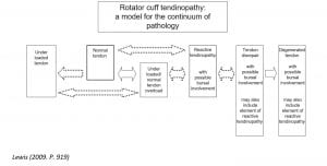
In the instance of rotator cuff muscle tendinopathy, the amount of load applied to the tendon is one of the most significant factor in the progression or regression of the pathology. As shown in the continuum below, when a normal tendon is not generally loaded properly, when intensity or frequency of load is increased suddenly, this may lead to overloading of the tendon and subsequent reactional tendinopathy. Similarly, when a normal loaded tendon is subject to a relative increase in load frequency and/or intensity, this may also lead to reactive tendinopathy. Whether the tendon is adequately loaded prior to excessive overload or not, the outcome is the same and results in reactive tendinopathy.
If this overload is not addressed and decreased, then further degeneration can occur and the condition goes beyond a sub-acute nature to a condition known as tendon disrepair, resulting in swelling and structural changes to the tendon, such as separation of collagen fibres which eventually leads to degeneration of the tendon, resulting in glenohumeral joint degeneration and large partial, full or massive thickness tears ( Lewis & Lewis, 2015).

According to Lewis (2009), the main goals in tendinopathy rehabilitation are load control, pain management, preventing further injury, patient education.
Based on the model created by Lewis (2009) and as shown in the following image:

and in the following link (I couldn’t upload a quality photo of the model) https://bjsm.bmj.com/content/44/13/918, loads placed upon the tendon, whether too much or too little, can have an effect on functioning ability and is a risk factor for tendinopathy.
Load reduction for reactive tendinopathy, therefore is paramount in preventing further degeneration, however load is still required in order to allow the tendon to increase capacity to withstand further loading.
It is recommended that load is increased gradually and within the capacity of the individual, so as not to irritate any acute inflammation or cause further regression in function and pain;
isometric, concentric and eccentric exercises are advised and as such the following exercises were given, targeting the affected tendons;
Isometric bicep contractions using resistance band 3-4 sets x15-40seconds
Isometric external rotation at 90 degrees 3-4sets x 15-40secs
Closed kinetic chain exercises for flexion and abduction by table slides 2-3sets x 12-14 reps.

I also took this patient’s posture into consideration; he had a slight kyphosis of the thoracic spine and as widely reported, this can contrite to upper body pain and dysfunction.
It is suggested that increases in thoracic kyphosis can have an effect further down the kinetic chain, for example it can cause downward rotation and protraction of the scapular which can in turn compress the sub acromial bursae or rotator cuff tendons (Lewis and Valentine, 2010). This was further supported by Seitz et al. (2011) who listed glenohumeral kinematics and scapular alignment as significant external risk factors for shoulder tendinitis, as well as external and internal impingement. This is just a small incite into the association between postural abnormalities and shoulder pathology, but worth considering in this case. We educated the patient on posture and gave appropriate stretching and advise on how to improve this. I have a lecture on shoulder rehabilitation approaching, which will delve far deeper into the kinetic chain models and muscle firing patterns of the upper body and limbs, which will hopefully provide me with far more understanding on the mechanisms and risk factors in the development of shoulder injuries and pain.

The information I found regarding rotator cuff tendinopathy was found in my second hour in this clinic session from internet research, to detailed discussions with Mike Prynn and two other students; speaking with others about these theories are very helpful and although I feel as though I lose confidence, comparing my knowledge to others’, I feel as though it highlights my gaps in knowledge, so I can go away and conduct further research.

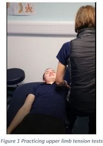
Patient 2 – My patient did not attend this session and fortunately there were other students in the clinic who also had time between appointments, so I took the opportunity to practice the upper limb neurodynamic tests.
In shadowing the previous treatment, I observed the application of a number of neurodynamic tests and although I had previously spent a significant amount of time researching the sensitivity and specificity of these tests and also watching a number of videos of how to perform them, I felt completely unable to carry out the tests and was unable to contribute to the session and instead just watched. I approached the therapist who I shadowed and asked him to go through the tests with me so that I was fully able to appreciate the exact procedure and he was kind enough to go through these with me.
The movements involved in this testing must be accurate as slight variations can change the test outcome; for example, incorporating scapular depression or blocking the scapular movement can result in either an earlier or later neurological response respectively (Legakis & Boyd, 2012).
I then spent time with fellow therapy students trying to navigate my way through the steps. I find it very difficult practicing provocation tests on healthy individuals who would not reproduce any symptoms and therefore provide positive findings for me to identify. However, I was able to identify if I was applying the correct movements and limb positions by the nature of nerve vs muscular capacity; if, during a ULNT a stretch is felt in the limb musculature for example, if I ask the individual to side bend their neck contralaterally, the ROM and sensation should be the same as before the neck same sensation. My understanding of this is that by adding the side bending of the neck to the contralateral side the nerves are put on more stretch and as such would inhibit the limb movements more than without the side bending. If no change is noted with the additional side bend, no cervical neuropathy is suspected.

The photograph in figure 1, is of me practicing the ulna nerve, although the photographer obviously did not capture the most exciting shot. Of all of the tests, I found this to be the hardest and could not replicate the movements as shown in the video I followed, which was as follows;
https://www.youtube.com/watch?v=rir6x6Iiqc4
After a few attempts, I decided to try to on another student, as I wondered whether this students hypermobility and flexibility might have influenced my inability to produce the expected stretch sensation in her forearm. This is an interesting point for discussion; was I just unable to perform the test properly, or does hypermobility and flexibility in a patient affect the ability to perform this test on an individual asymptomatic of upper limb neuropathy. My assumption is that this test would be physically possible if there was a positive test, as the nerve would produce the movement and pain restrictions instead of the muscular components of the arm.

My inability to perform these tests highlights the importance of physically practicing any practical elements of physical therapy and that watching videos alone, although very informative, are not enough to develop my practical skills. I can use the videos as a visual guide to go through the procedure step by step but I must practice this on other students before being able to perform them on live patients.

Patient 3 – Follow up for shoulder. Reported excellent progression with completely pain free motions and full ROM.
I believe the excellent progress can be credited to the compliance of the prescribed exercises; this patient was vey keen to return back to his recreational sports and as such fully adhered to the program we prescribed. Although he had a slight setback, due to trying to progress too quickly at one stage, by regressing the exercises we were able to start making progress again. I was really pleased that this patient returned to the clinic and to see the progress of this patient.
Unfortunately, however, as a result of occupational lifting, he sustained a back injury since his last treatment. This back injury led to complete inactivity for up to five days and the pain was described as agonising. Although this pain is greatly improved, the patient wanted to receive some advice on how to strengthen these now weakened structures. The patient also requested advice on a previous groin strain.
I performed an assessment on the patient’s back, from which I determined probable weaknesses in the gluteal muscles, particularly on the right side.
For the patient’s back pain, I prescribed some core stability exercises, glute strengthening as well as lumbar mobility exercises as these were all found to be effective in the treatment of lower back pain (Chang, Lin and Lai, 2015; Education, Gasibat, & Simbak, 2017; Purepong, Jitvimonrat, Boonyong, Thaveeratitham, 2012).
During the objective assessment it was clear that the patient’s pain was now localised to the right gluteal muscles and that the patient was no longer feeling any of the back pain or discomfort that disabled him two weeks prior to this session.
It was also observed that the patient had a slightly anteriorly tilted pelvis and as such I prescribed glute bridges with added isometric abduction exercises using a resistance band around the thighs (Choi et al., 2015).
From researching the effects of gluteal strengthening for the treatment of lower back pain, I came across a very useful article by Gasibat and Suwehli (2017), which reported positive effects of a number of exercises isolating the glutes and surrounding musculature.
This article also highlights the link between gluteal strength and lower back pain; any inactivity or guarding of the lower back during acute bouts of pain can lead to weaknesses in the gluteal muscles and therefore by strengthening these muscles, the risk of any changes in muscle activity due to pain and the evading of pain reduces (Gasibat & Suwehli, 2017).
One of the exercises that was advocated was a side plank for hip abduction, as shown in figure 2.

This would not only strengthen his abductors, including Gluteus Medius muscles, but his core muscles too. In order to add in some strengthening to the adductors too, to ensure that he can start running again soon without the apprehension of pain in the groin, we added resistance to this exercise by way of a chair. The up foot was to be rested on the chair while the lower leg is kept straight and raised off of the floor. This exercise can help to strengthen multiple muscle groups of which require attention in this particular patient.

In my reflections in the previous week, I wrote about the benefits and usefulness of squats and deadlifts in the strengthening of the lower extremity muscle groups and how I had hoped to be able to incorporate these into my clinic sessions and practice my ability to teach the correct technique to a patient.
I was very pleased to be able to use something new that I had recently learnt in this session and although weightlifting and any form of strength and conditioning is a new realm of knowledge for me, I knew that I would only learn by doing.
For this patient, deadlifts were prescribed with the aim of strengthening the erector spinae muscles (Schellenberg, Taylor and Lorenzetti, 2015) and as he does not have any access to a gym, we suggested using a backpack, within which he can add more weights if needed.
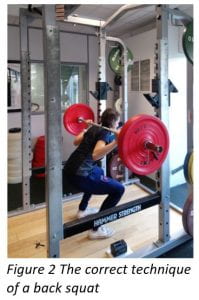
As he also presented with low level pain in his adductor muscles, I suggested incorporating back squats into his routine as well, but as recommended by Schoenfeld (2010) I remembered to adapt the squat, making sure the patient perform these with a wider stance.

Patient 4 – This session was a follow up session for a condition known as Scheuermann’s Kyphosis. The patient reported improvements in pain presentation from previous sessions of anteroposterior mobilisations of the thoracic spine, stretching of the pectoralis muscles, thoracic spine soft tissue manipulation and mobilisations. The patient also felt that postural Elastic Therapeutic Taping (ETT) was useful, as it provided the patient with a physical reminder to correct his posture by way of mechanoreception. I didn’t know much about this condition, so I took the time to develop my knowledge, should I encounter this condition again in the future.
According to a paper by Loughenbury & Tsirikos (2017), Kyphosis is now a disease believed to be the result of inheritance but often the cause is thought to be largely unknown (Sardar, Ames and Lenke, 2019) and presents itself as hyper kyphosis most usually in the thoracic spine and is diagnosed by an x-ray identifying a thoracic curve of 20-45° and other objective factors such as vertebral wedging, the narrowing between intervertebral discs and deformities in the effected vertebrae themselves. The wedging would be classified within the realms of this disease if it is of more than 5° of anterior wedging in three or more vertebrae (Sardar et al., 2019). The condition occurs in up to 10% of the population (Sardar et al., 2019) and causes pain and an obvious cosmetic deformity by way of an exaggerated curve of the thoracic spine, as shown in figure 3 (Loughenbury & Tsirikos, 2017).

The aforementioned studies highlight the fact that much of this condition is unknown, including how and why it occurs and as such, there is no clear consensus over treatment options, however it is reported that conservative treatment is effective and exercises can prove valuable in symptom management and inhibiting progression by way of strengthening the trunk muscles to improve balance and posture, increasing flexibility in the hamstrings and improving neck and shoulder mobility (Loughenbury & Tsirikos, 2017).
This is the same approach to the rehabilitation plan prescribed for this patient and so it will prove very interesting to watch his progression over the coming weeks with regards to pain and mobility throughout his spine and every day functional movements.

My final hour – I spent this time catching up on my clinic notes and discussing the treatments with the manager as a way of verbally evaluating and reflection on my performance as a therapist, as I was unable to complete this during my treatments; I still run over my session times and end up having to catch up by the end of the night. Due to the time of finish, it is within our duties to tidy and clean up the clinic before we leave. I feel as though we are becoming more proficient at this at the end of each session and are still maintaining good levels of cleanliness as seen at the start of the year.

References – 

Chang, W. D., Lin, H. Y., & Lai, P. T. (2015). Core strength training for patients with chronic low back pain. Journal of Physical Therapy Science, 27(3), 619–622. https://doi.org/10.1589/jpts.27.619

Choi, S. A., Cynn, H. S., Yi, C. H., Kwon, O. Y., Yoon, T. L., Choi, W. J., & Lee, J. H. (2015). Isometric hip abduction using a Thera-Band alters gluteus maximus muscle activity and the anterior pelvic tilt angle during bridging exercise. Journal of Electromyography and Kinesiology, 25(2), 310–315. https://doi.org/10.1016/j.jelekin.2014.09.005

Gasibat, Q., & Suwehli, W. (2017). Determining the Benefits of Massage Mechanisms: A Review of Literature. Article in Journal of Rehabilitation Sciences, 2(3), 58–67. https://doi.org/10.11648/j.rs.20170203.12

Legakis, A., & Boyd, B. S. (2012). The influence of scapular depression on upper limb neurodynamic test responses. Journal of Manual and Manipulative Therapy. https://doi.org/10.1179/2042618611Y.0000000020

Lewis, J. S. (2009). Rotator cuff tendinopathy/subacromial impingement syndrome: Is it time for a new method of assessment? British Journal of Sports Medicine, 43(4), 259–264. https://doi.org/10.1136/bjsm.2008.052183

Lewis, Jeremy S., & Valentine, R. E. (2010). Clinical measurement of the thoracic kyphosis. A study of the intra-rater reliability in subjects with and without shoulder pain. BMC Musculoskeletal Disorders. https://doi.org/10.1186/1471-2474-11-39

Lewis, Jeremy S, & Lewis, J. S. (2015). Rotator cuff tendinopathy : A model for the continuum of pathology and related management Rotator cuff tendinopathy : a model for the continuum of pathology and related management. Br J Sports Med. https://doi.org/10.1136/bjsm.2008.054817

Littlewood, C., Malliaras, P., Bateman, M., Stace, R., May, S., & Walters, S. (2013). The central nervous system – An additional consideration in ‘rotator cuff tendinopathy’ and a potential basis for understanding response to loaded therapeutic exercise. Manual Therapy. https://doi.org/10.1016/j.math.2013.07.005

Loughenbury, P. R., & Tsirikos, A. I. (2017). Scheuermann’s kyphosis: diagnosis, presentation and treatment. Orthopaedics and Trauma, 31(6), 388–394. https://doi.org/10.1016/j.mporth.2017.09.010

Purepong, N., Jitvimonrat, A., Boonyong, S., Thaveeratitham, P., & Pensri, P. (2012). Effect of flexibility exercise on lumbar angle: A study among non-specific low back pain patients. Journal of Bodywork and Movement Therapies, 16(2), 236–243. https://doi.org/10.1016/j.jbmt.2011.08.001

Sardar, Z. M., Ames, R. J., & Lenke, L. (2019). Scheuermann’s Kyphosis: Diagnosis, Management, and Selecting Fusion Levels. The Journal of the American Academy of Orthopaedic Surgeons. https://doi.org/10.5435/JAAOS-D-17-00748

Schellenberg, F., Taylor, W. R. ., & Lorenzetti, S. (2015). Exercise specific loading conditions and movements of squats, lunges, goodmornings and deadlifts. 33rd International Conference on Biomechanics in Sports.

Schoenfeld, B. J. (2010). Squatting kinematics and kinetics and their application to exercise performance. Journal of Strength and Conditioning Research.

Seitz, A. L., McClure, P. W., Finucane, S., Boardman, N. D., & Michener, L. A. (2011). Mechanisms of rotator cuff tendinopathy: Intrinsic, extrinsic, or both? Clinical Biomechanics, 26(1), 1–12. https://doi.org/10.1016/j.clinbiomech.2010.08.001

Exmoor Osteopathy External Placement Thursday 21st November 2019 – 4 hours (09:00-13:00): 121.5 total

Running total of hours: 121.5

During this session, had the opportunity to carry out two initial diagnostic assessments alongside my supervisor. Before I started this academic year, my weakest area of knowledge and practical ability was by far the vertebral column, so taking up an opportunity to work for Exmoor Osteopathy was a very positive decision in the development of this area in my knowledge. The number of patients who walk through the door with back pain is vast and as such, I have been able to experience various different presentations of a wide range of spinal dysfunctions, injuries and pathologies.
According to Gordon & Bloxham (2016), between 60 and 80% of adults will have lower back pain in their lifetimes, a condition which is responsible for 12.5% of work absences and costs the NHS £1.3 million each year.
The lower back pain patients attending this clinic are usually presenting with non-specific pain, which is thought to make up 85% of all back-pain cases (Gordon & Bloxham, 2016).

Throughout the treatments of LBP in this particular clinic, conservative passive treatments such as soft tissue and joint mobilisations, manipulations and massage are most commonly performed and are evidence based to some extent, but I try to offer my thoughts on including additional rehabilitation strategies such as stretching and strengthening, as well as patient education in order to allow the patient the opportunity to manage their lower back pain more independently and without the need for continual appointments at the clinic.
Throughout my reflections I have cited many studies that provide a large body of evidence suggesting that mobilisations, manipulations and massage etc are useful in reducing pain, improving flexibility and function, however these results are often short term or have not been proved to provide benefits that are beyond transient and as such more long term exercise programs are now more commonly recommended.

In a review by Gordon & Bloxham (2016), it was concluded that exercise is beneficial for individuals suffering lower back pain, but that no one exercise was better than the other; each case should be tailored for individually and should include strengthening, stretching and cardiovascular fitness as lower back pain can be reduced by up to 76.8% by core strengthening, 20% by cardiovascular fitness at 40-60% of heart rate reserve and 58% by flexibility training of the lumbar flexor and extensor muscles.

From reading this particular review, I was interested to learn the basic role of cardiovascular exercise in the management of pain; it is thought to stimulate the production of endorphins, which alters pain perception by way of binding to opiate receptors in the brain and spinal cord. Exercise also increases blood flow and nutrients to around the body and thus to the injury sites, which enhances the healing process by way of increasing mobility and enhancing the healing process (Gordon & Bloxham, 2016).
I am always an advocate for exercise of any form, so this was another incite as to the physiology behind it and another way I can help to explain on a basic level and reinforce physical activity in my future patients who may not be as physically active as they perhaps should be.

I also came across a randomised controlled clinical trial of 109 subjects, which found kinesiology taping effective in reducing pain in non-specific lower back pain (Kelle, Güzel and Sakalll, 2016). Whether this is down to a placebo effect or not and knowing the role of perception in pain presentation, a study like this is very useful in developing a basic level of confidence in this modality and I would personally feel more comfortable suggesting this treatment in future, to facilitate further exercise rehabilitation programs.

Although I enjoyed going through the assessment process for back pain and identifying possible treatment options based on each individual presentation, as I now feel more confident approaching this, the highlight in this session was our final patient attending for a follow up appointment about her knee.
Patient overview – this patient came to the clinic with knee pain and pain radiating down the lateral aspect of her lower limb (as far as 1/3 down the leg following the course of the peroneals). This was most likely degenerative and is now causing pain in functional, every day movements. This patient is over 60 but an active walker.
Conservative treatment for the past 2 months has not been as effective as hoped; soft tissue massage, mobilisations and stretching exercises were all performed but the patient only reported minimal improvements, with little difference in pain experienced in functional movements.
Katarina, the supervising osteopath discussed the possibility of a meniscal tear and the prospect of a referral for surgery. In many areas of musculoskeletal therapy, surgery is often avoided as much as possible and conservative treatment is our most favoured option with many soft tissue injuries due to the cost of surgery, the fear of longer rehabilitation times and the adverse associated risks of surgery. However, evidence suggests that in some injuries, the earlier the surgical intervention, the better the outcome of the injury in relation to the delays in future pathology. For example, with a meniscus tear, if surgery is performed early and is successful, future degeneration can be delayed and the occurrence of osteoarthritis of the knee comes later than if surgery is not performed and as such further inevitable surgery, such as a knee replacement is put off for more longer (Vaquero & Forriol, 2016).
In order to fully assess the knee and to diagnose a meniscus injury requiring surgery, this patient would need to undergo magnetic resonance imagining (MRI), which according to Vaquero & Forriol (2016) is 90.5% sensitive and 89.5% specific or an arthroscopy which is used to both diagnose and treat via a keyhole surgical method.
The location and degree of the tear can vary the surgical treatment and there are many contraindicators that can also restrict surgical outcomes and therefore surgical intervention is not always the most suitable treatment. The cost and time involved in a referral for the patient can be extensive and as such Katarina would not suggest this lightly. The purpose of further investigations would be to identify the need for surgery or to rule out anything that may require surgery, in order to fully trust the route of conservative treatment.
A study by Kise et al. (2016), comparing the outcome of physical therapy vs. arthroscopic partial meniscectomy found no significant differences between the two options, however thigh muscle strength was found to have improved more in the conservative approach and therefore the non-surgical route is recommended, supporting earlier findings by Stensrud, Risberg and Roos (2015) in a study showing improvements in isokinetic quadriceps strength after a 12-week exercise program for the treatment of degenerative meniscus tears.
Knowing the specific criteria for surgery and effectiveness of physical therapy programs, it would usually be appropriate to continue with strengthening, however because this individual patient was not experiencing any benefits from the physical therapy, it was worth suggesting a referral.
If surgery is not indicated, we will know that we can continue to treat this patient in a conservative manor, but unlike before the referral, I will hope to add a more strengthening heavy exercise plan, rather than the more passive treatment previously administered as this provided the patient with minimal strengthening rehabilitation.

References –

Gordon, R., & Bloxham, S. (2016). A Systematic Review of the Effects of Exercise and Physical Activity on Non-Specific Chronic Low Back Pain. Healthcare, 4(2), 22. https://doi.org/10.3390/healthcare4020022

Kelle, B., Güzel, R., & Sakalll, H. (2016). The effect of Kinesio taping application for acute non-specific low back pain: A randomized controlled clinical trial. Clinical Rehabilitation. https://doi.org/10.1177/0269215515603218

Kise, N. J., Risberg, M. A., Stensrud, S., Ranstam, J., Engebretsen, L., & Roos, E. M. (2016). Exercise therapy versus arthroscopic partial meniscectomy for degenerative meniscal tear in middle aged patients: Randomised controlled trial with two year follow-up. BMJ (Online). https://doi.org/10.1136/bmj.i3740

Stensrud, S., Risberg, M. A., & Roos, E. M. (2015). Effect of exercise therapy compared with arthroscopic surgery on knee muscle strength and functional performance in middle-aged patients with degenerative meniscus tears. American Journal of Physical Medicine and Rehabilitation. https://doi.org/10.1097/PHM.0000000000000209

Vaquero, J., & Forriol, F. (2016). Meniscus tear surgery and meniscus replacement. Muscles, Ligaments and Tendons Journal. https://doi.org/10.11138/mltj/2016.6.1.071


Clinical Experience Tuesday 19th November 2019 – 5.5hours (15:00-20:30): 117.5 total

Running total of hours: 117.5

Patient 1 – Antigravity Machine Follow – up I set up myself yay!
Patient Overview of three sessions beginning October 2019
Session 1 –Single-leg squat difficult and reproduces Px, twisting causes Px
Thessaly’s test, did not complete as single leg squat reproduces Px
Session 2 – Px in rot. Of knee during weight baring and PROM.
Session 3 – Px free for 2/52, now only 1/10 Px when twisting (at worst) but mostly Px free.
Excellent progress, completing exercises. Full active, passive ROM, 5/5 RROM. Single leg squat better but still not completely stable.

Although this patient has seen excellent improvement over the past three sessions, he is still experiencing instability during his more functional tasks, such as a single leg squat or similar. This was an exercise prescribed to this patient early on in his rehabilitation, yet it has not seemed to improve at the same rate as his pain. Because the patient was not able to perform this in a controlled manner, I did not progress his exercise routine, for example by adding resistance.

Deficits in proprioception can be a risk factor in musculoskeletal injuries and can result from muscle fatigue, trauma or gradual onset from chronic pain conditions (Röijezon et al., 2015). Proprioception is defined as an individual’s ability to integrate the sensory signals from mechanoreceptors to thereby determine body segment positions and movements in space” (Han, Waddington, Adams, Anson and Liu, 2016. p. 81).

While short term treatments such as massage, mobilisations, manipulations and taping can provide short term improvements in proprioception, for long term sustainable improvements in balance, the following exercise could be performed as recommended by Clark, Röijezon and Treleaven (2015); standing with eyes open on soft surface with a narrow tandem stance and performing a single leg squat, with progression opportunities for this whereby subject can close eyes, stand on uneven surface and add an external stimulus via a specific task such as throwing and catching a ball or performing this task on a rocker or wobble board as shown in figure 1.

This exercise can be used as a method of testing the patient’s proprioception by providing a good objective measure; how long can the patient perform this task before fatiguing or losing control (Clark et al., 2015).

Although it is reported that there is a lack of randomised clinical trials to support the incorporation of proprioception training in the treatment of OA, it is still recommended as a means to reduce pain and function (Nguyen, Lefèvre-Colau, Poiraudeau, Rannou, 2016).
A study on 40 subjects with knee osteoarthritis by Lai, Mason, Tan and Boyle (2018) found that knee joint motion sense during flexion was significantly enhanced after an 8week intervention training program of squats.

Topp & Pifer (2017) discussed the effects of knee AO on knee position sense and proprioception, stating that subjects with knee OA experience reduced proprioception in the affected knee and after conducting a study on the effects of different proprioceptive training methods, found that a 16 week program of resistance band isometric exercises was the most effective method to reduce unilateral deficits by way of strengthening the supporting structures and subsequently improving neurological stimulation and coordination during functional movements. Dynamic resistance training was also found to be of similar benefit to isometric resistance training and as such was also recommended as an intervention in knee OA (Topp & Pifer, 2017).

The balance task that was prescribed for this patient will hopefully further improve pain and function in his knee, enabling us to progress further into a more multifunctional approach and incorporate more dynamic movements involving twisting and turning on the knee; the mechanism in which reproduces the pain the most.

Patient 2 –
This patient was a young male runner who presented with pain in his posterior leg, slightly inferior to his knee. After an assessment whereby a full knee assessment was performed, the clinic supervisor needed to come to my rescue; I had never come across posterior knee pain before and my knowledge of the anatomy here was very limited.
The patient experienced pain only in functional movements and occasional , so it was very difficult to reproduce the same pain in passive, active or resisted movements. The only movement that slightly reproduced the pain was resisted inversion and resisted plantarflexion, but with no compromised neuromuscular strength. Palpation and running produced pain behind his knee.
The clinic supervisor was unsure as to the exact location of the pain, however the nature of the pain, e.g. feeling of ‘tightness’, pattern of pain; worse in the morning, eases during the day and through exercise was indicative of a tendinopathy of one of the tendons of the posterior knee.
Of the posterior structures of the knee, both the popliteal tendon and the plantaris tendon were considered as the source of pain in this instance, however popliteal tendinopathy is regarded as a rare condition (Doucet, Gotra, Reddy and Boily, 2017) and due to the mild presentation of symptoms and that there was no reproduction of symptoms in flexion and rotation of the knee, the actions produced by the popliteus, we were unable to confirm the involvement of this muscle. Conversely, the plantaris both plantarflexes and inverts the foot, both of these actions reproducing the pain for this patient, therefore allowing us the assumption that this is the source of the patient’s tendinopathy type symptoms.

In my research into this condition, I discovered the relationship between achillies tendinopathy and the plantaris muscle. The plantaris muscle, as shown in figure 2, is often reported as a contributor to tendinopathy of the achillies tendon although it is unclear as to how. It has been suggested that the plantaris muscle’s location, in relation to the other muscles in the lower leg (the achillies tendon, tibialis posterior) could be a factor, as the plantaris location varies between individuals (Olewnik, Wysiadecki, Podgórski, Polguj and Topol, 2018).

The interesting aspect of this patient’s injury is the history of achillies tendinopathy over six months prior to this treatment and although the two conditions are seemingly separate, the two injuries may actually be related or have somehow been misdiagnosed. According to Alfredson, Masci and Spang (2017), mid-portion achillies tendon pain may actually be plantaris tendon pathology but with similar symptoms and is therefore overlooked. This poses the question of whether the patient’s previous injury was misdiagnosed as non-insertional achillies tendinopathy or whether this injury has provoked further inflammation of the plantaris muscle. Koh, Mason, Tan, Boyle (2018) reports that the plantaris muscle could also be a possible contributor to achillies tendon injuries, so it may remain unknown as to the development of this particular injury.
Although we had an idea as to the exact location or cause of the injury, we were unable to give a definitive diagnosis due to the mild nature of the patient’s symptoms and the aforementioned relationship between the achillies and plantaris tendons.
As the two conditions are similar in presentation, it was tempting to prescribe a training plan similar to rehabilitation of the achillies tendon. As mentioned by Koh et al. (2018), however, it is important not to perform eccentric exercises in this instance, as this would worsen symptoms by way of increasing friction and therefore inflammation.
Isometric exercises can be performed to reduce pains symptoms (Koh et al., 2018) but initially, exercises used in rehabilitation for acute tendinitis would be more appropriate as this would aim to reduce the friction and inflammation and reduce loading to a minimum which reduces the likelihood of developing into degenerative tendinosis.
In some cases, surgery to remove the plantaris tendon is reported as a successful procedure (Alfredson et al., 2018), which further highlights the role of the plantaris in achillies tendon injuries. Without diagnostic ultrasound or other more details means of radiography, our goal of treatment was to reduce pain and increase function in this patient and so strengthening exercises, targeted at the plantaris were prescribed but at low intensity so as not to irritate the tendon and surrounding structures.

Patient 3 – I did not have a patient in this hour of my clinic session, but I used this time to catch up on clinic notes and research the pathologies from the previous sessions, preparing for their return appointments. I find it especially useful conducting the research immediately post clinic or patient as it is fresh in my mind. I also feel that it is very important to prepare for the next appointment well in advance, in case they move their appointments forward last minute and also due to the fresh information in my mind.

Patient 4 and 5 – Two of my patients in this session presented with shoulder pain, one of the patients being a middle-aged male with a very active job and the other being an elderly male who is soon to retire from an equally physical occupation. Both patients were physically active and their assessments were very similar in nature.

Earlier in my clinical experiences, on Monday 4th November, I reflected upon a treatment I did on a patient with rotator cuff pathology.
Within the reflections, I had commented on the level of rehabilitation that I prescribed my patient and the lack of physical demand; the patient was already a fit, active individual who played good level of tennis on a regular basis and as such, the exercises I prescribed may not have been challenging enough.
Unfortunately, I have not had the chance to see this patient since her initial appointment (either she felt that the rehabilitation plan given was sufficient in facilitating her own progress back to full functional ability, or that she did not feel as though the rehabilitation was helpful). I had hoped to monitor her progress by reassessing pain and the time of onset of pain since her initial appointment so that I could prescribe a more suitable plan.

From the failings of this session, I have better understood the need to individualise each program and take into account the patient’s current levels of activity.
In these instances, both patients were physically active and had occupations that required heavy lifting, therefore their need to incorporate strength specific training may be less than the need to increase endurance.
As mentioned earlier in my reflections, recommendations by Ellenbecker & Cools (2010) to include resistance training exercises are there to invoke a fatiguing response to the exercises and thus increase endurance. In this case, both patients may have good strength in their shoulders, as indicated in their objective assessments, but the time frame of onset of pain during their daily activities are indicative of weakness in the muscles, possibly due to early fatigue. I utilised their recommendations of fifteen to twenty repetitions of three sets as this would increase strength endurance rather than strength capacity. Any initial progression in resistance would be of repetitions and sets but not necessarily the weight.
Interestingly, my reflections highlighted the differences between the amount of loading placed upon the resistance movements, stating that higher loads activate the deltoids, but lower loads activate the supraspinatus Ellenbecker & Cools (2010).
In addition to this, I have since learnt that in order to isolate separate muscle groups, as well as varying the load, it is the angle in which the exercise is performed; when the arm is being worked at the end range of abduction, this activates the deltoids but it the exercises are being performed at the start range and more specifically in the scapular plane, this activates the supraspinatus, subscapularis and infraspinatus (Reed, Cathers, Halaki and Ginn, 2016).
It is important to prescribe exercises specific to the affected muscle, so as not to allow the recruitment and activation of the more dominant muscles within the shoulder. The recruitment of muscles used in each movement of the shoulder is complex with a vast assortment of muscles activated to facilitate and stabilise movements, but when there is weakness and pain in some muscles other structures may overcompensate.

Usually in the instance of rotator cuff tendinopathy, exercises to adequately load the tendons are most appropriate in order to increase ROM and reduce pain (Lewis & Lewis, 2015). However, I have since attended a very informative lecture, within which the whole shoulder and associated kinetic chain was discussed in a whole new light to me; models suggestive of other muscle group involvements and dysfunctions led me to question my previously assumed tendon pain in the subjects presenting with other symptoms, specifically posture related.
In my previous sessions with patients presenting in shoulder pain whereby tendinopathy is most likely, I have not considered the possible extrinsic risk factors involved and merely treated the symptoms of tendinopathy, assuming biomechanical dysfunction resulted solely from increase in load. As reported by Seitz, McClure, Finucane, Boardman and Michener (2011), however other external risk factors involving shoulder kinematics and posture could have a role in the development of in injuries such as tendinopathies or external and internal impingement. With both intrinsic and extrinsic factors possibly posing a risk for shoulder pain and the multitude of possible causes of pain, Lewis (2009) proposed the term mechanical shoulder pain, as determining an exact cause or definition may not be possible.
According to Cools et al. (2014) muscle weakness, lack of flexibility and thoracic curvature can all be external factors in the development of pain and as such exercises to target these specific dysfunctions are important.
Stretching the pectoralis muscles using the door frame stretch is something I use regularly for my patients, but the sleeper stretch, as used by Cools et al. (2014) is a new option for me to use.
I have a regular patient who could benefit very much from pectoralis stretching, but she cannot perform the door frame stretch due to lack of mobility, pain and range of motion in the shoulder and so I have not been able to incorporate pec stretching in my sessions but now, having come across these recommendations, I can prescribe a stretch that the patient can mostly definitely perform as it required the patient to lie down; she can do this before she gets out of bed in the mornings or at night before sleep.
In order to strengthen the muscles that stabilise the scapular, Cools et al. (2014) suggested targeting thoracic spinal muscles such as the lower traps, rhomboids and serratus anterior with exercises such as the lawn mower, inferior glide, low row and robbery.
Closed chain exercises were also suggested as a way to stabilise the scapular as it allows the patients to focus on where the scapular is in relation to their chest wall and activate muscles solely responsible for stabilising the scapular as apposed to global movers within the shoulder, with further examples of those I can aim to use in the future shown in the following video. I can look to develop these within the clinic.

Scapular dyskinesia exercises –
https://www.youtube.com/watch?v=FBJHyT_MtzA&feature=youtu.be

In order to fully understand the possible involvement of kinematics and biomechanical influences on shoulder pain, there are various different methods of assessment, one being the Shoulder Symptom Modification Procedure (V2), originally designed by Lewis (2009), found in the following document.

SSMP- Jeremy Lewis V2

For pain under the scope of mechanical shoulder pain, as suggested by Lewis (2009) whether thoracic curvature, scapular position or dysfunction or humeral head position is the cause of the pain or shoulder injury, or whether another source of pain was the cause of the misaligned shoulder kinematics, methods to correct this can be incorporated into treatment. If, when assessing and working through the SSMP, any symptoms are relieved, then this is the assumed area of dysfunction and should therefore be the subject of treatment options.

From reading the research surrounding the shoulder and associated biomechanics, I have really developed my understanding on the bigger picture and the other factors that can be involved with but also managed. With the lack of testing procedures of high specificity or sensitivity, it is useful to have another route of investigations to find perhaps a more specific answer as to how the pain has occurred, especially in those patients who are assumed to have tendinopathy but who are not experiencing any improvements.

References – 

Alfredson, H., Masci, L., & Spang, C. (2018). Surgical plantaris tendon removal for patients with plantaris tendon-related pain only and a normal Achilles tendon: A case series. BMJ Open Sport and Exercise Medicine. https://doi.org/10.1136/bmjsem-2018-000462

Clark, N. C., Röijezon, U., & Treleaven, J. (2015). Proprioception in musculoskeletal rehabilitation. Part 2: Clinical assessment and intervention. Manual Therapy, 20(3), 378–387. https://doi.org/10.1016/j.math.2015.01.009

Cools, A. M. J., Struyf, F., De Mey, K., Maenhout, A., Castelein, B., & Cagnie, B. (2014). Rehabilitation of scapular dyskinesis: From the office worker to the elite overhead athlete. British Journal of Sports Medicine, 48(8), 692–697. https://doi.org/10.1136/bjsports-2013-092148

Doucet, C., Gotra, A., Reddy, S. M. V., & Boily, M. (2017). Acute calcific tendinopathy of the popliteus tendon: a rare case diagnosed using a multimodality imaging approach and treated conservatively. Skeletal Radiology. https://doi.org/10.1007/s00256-017-2623-8

Ellenbecker, T. S., & Cools, A. (2010). Rehabilitation of shoulder impingement syndrome and rotator cuff injuries: An evidence-based review. British Journal of Sports Medicine, 44(5), 319–327. https://doi.org/10.1136/bjsm.2009.058875

Han, J., Waddington, G., Adams, R., Anson, J., & Liu, Y. (2016). Assessing proprioception: A critical review of methods. Journal of Sport and Health Science, 5(1), 80–90. https://doi.org/10.1016/j.jshs.2014.10.004

Koh, E., Mason, M., Tan, S., & Boyle, J. (2018). Imaging of plantaris friction syndrome. Clinical Radiology. https://doi.org/10.1016/j.crad.2018.05.023

Lai, Z., Mason, M., Tan, S., & Boyle, L. (2018). Effects of strength exercise on the knee and ankle proprioception of individuals with knee osteoarthritis. Research in Sports Medicine. https://doi.org/10.1080/15438627.2018.1431541

Lewis, J. S. (2009). Rotator cuff tendinopathy/subacromial impingement syndrome: Is it time for a new method of assessment? British Journal of Sports Medicine, 43(4), 259–264. https://doi.org/10.1136/bjsm.2008.052183

Lewis, Jeremy S., & Valentine, R. E. (2010). Clinical measurement of the thoracic kyphosis. A study of the intra-rater reliability in subjects with and without shoulder pain. BMC Musculoskeletal Disorders. https://doi.org/10.1186/1471-2474-11-39

Lewis, Jeremy S, & Lewis, J. S. (2015). Rotator cuff tendinopathy : A model for the continuum of pathology and related management Rotator cuff tendinopathy : a model for the continuum of pathology and related management. Br J Sports Med. https://doi.org/10.1136/bjsm.2008.054817

Nguyen, C., Lefèvre-Colau, M. M., Poiraudeau, S., & Rannou, F. (2016). Rehabilitation (exercise and strength training) and osteoarthritis: A critical narrative review. Annals of Physical and Rehabilitation Medicine, 59(3), 190–195. https://doi.org/10.1016/j.rehab.2016.02.010

Olewnik, L., Wysiadecki, G., Podgórski, M., Polguj, M., & Topol, M. (2018). The Plantaris Muscle Tendon and Its Relationship with the Achilles Tendinopathy. BioMed Research International. https://doi.org/10.1155/2018/9623579

Reed, D., Cathers, I., Halaki, M., & Ginn, K. A. (2016). Does load influence shoulder muscle recruitment patterns during scapular plane abduction? Journal of Science and Medicine in Sport, 19(9), 755–760. https://doi.org/10.1016/j.jsams.2015.10.007

Seitz, A. L., McClure, P. W., Finucane, S., Boardman, N. D., & Michener, L. A. (2011). Mechanisms of rotator cuff tendinopathy: Intrinsic, extrinsic, or both? Clinical Biomechanics, 26(1), 1–12. https://doi.org/10.1016/j.clinbiomech.2010.08.001

Topp, R., & Pifer, M. (2017). A Preliminary Study Into the Effect of 2 Resistance Training Modes on Proprioception of Subjects with Knee Osteoarthritis. JPHR: Journal of Performance Health Research. https://doi.org/10.25036/jphr.2017.1.1.topp


Clinical Experience Monday 18th November 2019 – 4.5hours (16:00-20:30): 112 total

Running total of hours: 112

Patient 1 – STM of lower back to relieve sciatica and chronic LBP

Patient Overview: from a thorough assessment, our clinical impression was that of chronic LBP and muscular dysfunction of and around LX and Tx, weakness in hamstrings and quads as a result of slight anterior tilt of pelvis and subsequent LBP.

For this patient, our primary outcome was to reduce pain and increase strength mobility in his lumbar and thoracic spine in order to do so. I used pain and ROM as clinical markers for the treatment intervention, testing both before and after the treatment. Initially I used STM which has been found to be effective on chronic lower back pain (Kumar, Beaton and Hughes, 2013; Romanowski and Grześkowiak, 2012). As both PA grade IV mobilisations and prone lumbar extensions have been found to reduce pain and improve function in chronic lower back pain (Shah & Kage, 2016; Shum, Tsung and Lee, 2013; Varun, Manoj, Jaspreet and Naveen, 2014), I performed both of these treatments.
Although I am confident that I have found some supporting evidence on the effects of PA, I am not entirely sure as to the exact procedure involved in terms of intensity and duration.
According to Shah & Kage (2016), the mobilisations that brought about positive effects were as follows;
10 minute PA treatment in total, involving grade IV oscillations (or as pain guided by the individual, but must not reproduce symptoms) of lumbar vertebrae = 3 sets of 40seconds
Shum et al. (2013) found positive effects from 3 sets of grade III oscillations at the end range for up to 60seconds.

The 10minute prone press ups treatment in the same study (Shah & Kage, 2016) was performed by the patient moving into full, tolerable extension using arms to bring chest and upper body up with pelvis against the ground: 10 repetitions of 5 seconds each, increasing to 2 or 3 sets depending on pain. This is very similar to the McKenzie method which is used for the treatment of lumbar disc herniations.

I also included stretching and a prescription of exercises to strengthen the iliopsoas, back extensors, abdominal muscles and hamstrings as shown be effective by Kachanathu, Alenazi, Seif, Hafez and Alroumim (2014). A study by Chang, Lin and Lai (2015) also recommended the incorporation of core strengthening, particularly deeper structures namely the internal obliques, transversus abs, lumbar multifidus and quadratus lumborum muscles in a program to treat reduce LBP as it is often reported that weaknesses in these muscles contribute to CLBP.

Notes as recorded on the Cliniko Client Management Program

The patient overview is shown in the soap notes above. Over the past two months I have tried to use abbreviations where possible so that the notes can be universally read and more efficiently. I have found that in the past I write too much on the assessment forms, which takes up time in the consultation and proves difficult to read in the future; when trying to read follow up notes in future appointments, it is really difficult to decipher through the relevant information.
Using abbreviations took more time initially as I was getting used to what they all meant, however I am becoming more proficient at this during each session. There is still inconsistency in the notes above, for example I have still used the work “pain” instead of Px and full muscle names such as “hamstrings” and “quadriceps” where I could instead replace these with “hams” and “quads”. However, I am happy with my progress and hope to further develop my ability to type up short notes quickly but without compromising the detail.

Looking back on previous notes, it came to my attention that I was not writing full range of motions and only recording the ones of relevance and stating “Full A, P and R ROM”. I have since been asked to be more specific in this objective write up and instead of writing this, include which ROM tests I did. For example, Full Active Flex, Ext, Int. & Ext. Rot., Add., Abd., h.abd. and to include Lt and Rt. Although this is far more time consuming, it is important to be this specific so that any therapist reading these notes will know which movements have been tested and to be sure that no movements were forgotten and to know which movements were not tested.

It is also educational to read through previous notes on the patients to reflect back not only on their treatment progress but on my note writing and understanding of the patient and assessment as well as other student’s interpretation of results.
I had not seen this patient before, so previous notes were those written by other students. Although I write in a similar manner, using the same abbreviations as per the protocol in this clinic, I have been able to pick up a few descriptive words that I may not have used before. In this instance, the word “hypertonic” was used. Interestingly, I have struggled to find the correct terms for this as tension is not quite the right term.

With regards to the detail I add into each write up, I now incorporate more specific information about the exercises I prescribe, as I feel that this is an important aspect of an ongoing treatment plan. Knowing the repetitions and sets of certain exercises and exactly the design of the exercises are important.
For example, in this case the patient reported that his list of exercises were extensive and as such he felt as though he did not have enough time to complete them each day. It is important to discuss the rehabilitation on a regular basis with the patient, because if they do not feel as though they can complete the session, theur adherence may reduce. Adding too many exercises or increasing the intensity of the exercises before they are ready could effect their compliance.
Interestingly, however an early study by Perri et al. (2002) on the effects on changing frequency and intensity on exercise adherence, found that increasing the frequency did not reduce adherence but that increasing frequency did. Although this was a study on cardiovascular fitness, time constraints still have a role in completing these exercises, especially when frequency is increased. This highlights the needs to approach each patient on an individual basis.

It was reported that patient education and the ability to incorporate exercises into the daily routine of the patients were two of the most influencing factors contributing to patient adherence to exercise programs in physiotherapy (Bassett, 2015). It is our job, therefore as therapists to make the program as appealing as is possible and to spend plenty of time without the session to explain what is expected of them and ensure that they fully understand their rehabilitation plan, their goals and outcomes and make sure that both the patient and the therapist are in agreement with the plan.

In this case, the patient reported the sessions consisting of too many activities, so in order to break this down but without taking away any of the exercises, I suggested halving the load and completing each half on alternate days, which the patient seems much happier with.

Patient 2 – Undiagnosed Scoliosis
This patient was a young male who presented with chronic and undiagnosed back pain.
Just from an objective assessment, it became apparent early on that there was scoliosis in the distal portion of the thoracic spine. Scoliosis is a spinal condition whereby the spine deviates 3-dimensionally and exhibits curvature of around 10° in the coronal plane shown by an x-ray taken anterior posteriorly (Trobisch, Suess and Schwab, 2010). It is reported to affect up to 2% of children, 8% of adults and 68% in the elderly population over 60 (Trobisch et al., 2010).
In order to properly assess the condition and to discuss findings with the patient, we videoed the patient performing forward flexion using the clinic iPads. Assessing the curve in this functional movement has been reported as an effective means to test the extent of the deformity and the nature of the stiffness (Silva & Lenke, 2010). Interestingly, leg length is a risk factor for scoliosis and as such, inserts are a possible treatment intervention (Silva & Lenke, 2010), however I did not test for this in the first assessment and will be sure to test for this in the follow up.

HPC
13 years old back Px, no significant onset, intermittent Px around 2/10,
some good, some bad days. 2/52 ago agony. Aches and sharp stabbing Px
20-30mins at time.
Lay on floor until subsides – 9-10/10
Bending over causes onset
4 years ago GP, had physio. GP = scoliosis, physio = imbalance of muscles.
Left muscle bulk length of spine. Physio = nothing to be done.
No night Px
No Spasms
Can’t sit down for long periods of time
No Px down leg/nerve Px
At present 6/10
Used to play cricket and football but stopped due to Px
Heavy lifting all day so very active
Px is manageable at work.

O
Full active ROM flex, ext, bilat. Side flex and rot. No Px
ipsilateral side px during combined lx ext. And Rt Rot.
Jt clear above
Jt below (hip) no Px in all ROM but instability and hitching in left hip during Rt hip flex

Single leg stance – instability and hitching in contralateral hip during Rt hip flex.

As recorded in the assessment notes shown above, this patient has had this condition for 13 years and as this patient is still in his 20s, this suggests that it could have been diagnosed as adolescent or even childhood scoliosis. In adolescent with curves greater than 20°, progression is unlikely, however this is not true for each individual case. In individuals with curves greater than 30°, the deformity is likely to progress up to 1-1.5° per year
(Silva & Lenke, 2010; Weinstein et al., 2013).
In this case, as the patient reported symptoms appearing in his teenage years, suggestive of, and in the absence of a proper clinical diagnosis, adolescent scoliosis and as his curve does not exceed 20°, he has just a 10-20% chance of the deformity progressing.
With regards to treatment, according to Negrini et al. (2015), physical therapy including exercises to correct alignment and improve balance affected by the collapse in the contralateral side to the curve is an effective means to manage pain, but with surgery being the only known means to stop curve progression. Because of the abnormal spinal curvature, other structures in the back and shoulders have been compromised and as such our treatment plan focused solely on creating a more symmetrical movement pattern and alignment throughout his spine which will aim to reduce the pain the patient is currently experiencing.
When diagnosed early in juveniles or adolescents, treatment is tailored to prevent progression and where there is a severe curve, interventions such as surgery or bracing could be performed, with bracing having been shown to have significantly reduced the progression of greater curves, with a correlation between the time the brace is worn and the progression delay (Weinstein et al., 2013).

Although this patient is beyond his adolescent years, he is still relatively young and treatment to relieve symptoms can be performed and prescribed if only to reduce pain and improve function and not inhibit the progression of the condition. Earlier in my reflections I came across a patient who had scoliosis diagnosed in early life and I had hoped that I would find the time to research into this condition and possible treatment outcomes.
As the previous patient with scoliosis was no longer undergoing treatment for his spine alone, because he had already experienced huge changes in his posture and pain levels, it was difficult for me to visually appreciate the effects that the treatment had and the difference it made on the individual. There is little to no evidence to suggest the effectiveness in any non-surgical treatments to reduce the progression of scoliosis, however the patient seen in my external placement reported significant postural changes, even at an older age and as such I feel positive that a treatment intervention is worth undergoing.

I am excited to start a treatment plan for this patient and to observe changes, if any, that may come about.

Figure 1 is of the video that we took at the beginning of treatment. This is an effective way of objectively recording any differences throughout rehabilitation and also gave us the opportunity to show the patient what we saw as he repoerted that he had not seen his spine. The photos show the curavture of the spine to the right at the distal thoracic spine.

Extra Practice between Patients due to cancellations
Due to two cancellations, I had some time to gain more experiencing using the clinic equipment or develop knowledge on certain aspects of the clinic that I am not usually able to do due to the back-to-back nature of the clients. I find this time extremely important in familiarising myself with all areas in the clinic, especially those I feel less confident with.
During these first 3 months of clinic, I have not yet had the opportunity to use the strength and conditioning gym or equipment and I feel that this is my greater area of weakness. Unfortunately, however, this is one of the most important aspects of musculoskeletal injury treatment and prevention and is an effective way to enhance lower body strength, specifically of the leg, hip and back (Deforest, Cantrell, Schilling, 2014). It has also been suggested that due to the simplicity of squatting, it is an extremely effective exercise in improving quality of life; once movement elicits muscular responses from multiple muscle groups (Schoenfeld, 2010). The motion is also similar to that of many everyday tasks and as a tool for rehabilitation, could prove useful in encouraging exercise adherence, as incorporating exercises into everyday activities has been found to increase the likelihood of patients completing rehabilitation (Bassett, 2015), as mentioned earlier in my reflections. Due to the close-kinetic-chain nature of this exercise, it can be introduced early in rehabilitation programs and progression by way of weight resistance is not exhaustive (for the purpose of rehabilitation and injury prevention). Although the squat exercise creates little injury risk, when performed using the incorrect technique, problems can arise. It is also important to know about how variations of the squat technique can be made and adapted to suit the needs of the individual patient. For example, the depth and execution of the squat can determine where in the kinetic chain the load is most applied, which muscle groups are being worked the most and to reduce detrimental or damaging effects. According to Schoenfeld. (2010), the key aspects of the squat include injury site, speed of squat, depth of squat and stance; rehabilitation for injuries concerning the knee should not perform deep depth squats and must be performed with a wider stance, time of 2 to 3 seconds for a controlled eccentric motion is most efficient and safe to reduce loading on the joint and parallel squats are most effective to strengthen quadriceps whereas a wider stance is used to focus on hip adductors and extensors.

Schellenberg, Taylor and Lorenzetti,(2015) found that deadlifts and good mornings, as shown in the following video were found to effectively strengthen the erector spinae muscle group and as such would be a good exercise to prescribe for patients with lower back pain. This exercise can be done whether the patient has gym membership or not.
Providing the patients have the correct technique with the deadlift, instead of a barbell, household items can be used such as shopping bags and weight can be added inside the bag to increase resistance.
The correct technique for a Good Morning
https://www.youtube.com/watch?v=YA-h3n9L4YU
The correct technique for a Deadlift
https://www.youtube.com/watch?v=op9kVnSso6Q

These videos were excellent demonstrations of the squats, both of which I had never performed before. From having tried this, I am now able to better explain the technique involved and how it feels. I also learned very early on that the bar was very uncomfortable on my back and as such I added some bar specific padding, which enabled me to perform the squat without the discomfort. As a group of students, we practiced the aforementioned squats with figure 2 showing me performing the back squat.

This session proved a good introduction to the benefits of squatting on lower extremity injury prevention and strengthening, as my prior knowledge was next to none. I can hope to continue my knowledge on this aspect of rehabilitation and strength and conditioning throughout the relative module and by incorporating more strength and conditioning exercises and protocol in my treatment plans.

References –

Bassett, S. (2015). Bridging the intention-behaviour gap with behaviour change strategies for physiotherapy rehabilitation non-adherence. New Zealand Journal of Physiotherapy, 43(3), 105–111. https://doi.org/10.15619/nzjp/43.3.05

Chang, W. D., Lin, H. Y., & Lai, P. T. (2015). Core strength training for patients with chronic low back pain. Journal of Physical Therapy Science, 27(3), 619–622. https://doi.org/10.1589/jpts.27.619

Deforest, B. A., Cantrell, G. S., & Schilling, B. K. (2014). Muscle Activity in Single- vs. Double-Leg Squats. International Journal of Exercise Science, 7(4), 302–310. Retrieved from http://www.ncbi.nlm.nih.gov/pubmed/27182408%0Ahttp://www.pubmedcentral.nih.gov/articlerender.fcgi?Artid=PMC4831851

Kachanathu, S. J., Alenazi, A. M., Seif, H. E., Hafez, A. R., & Alroumim, A. M. (2014). Comparison between Kinesio taping and a traditional physical therapy program in treatment of nonspecific low back pain. Journal of Physical Therapy Science, 26(8), 1185–1188. https://doi.org/10.1589/jpts.26.1185

Kumar, S., Beaton, K., & Hughes, T. (2013). The effectiveness of massage therapy for the treatment of nonspecific low back pain: A systematic review of systematic reviews. International Journal of General Medicine. https://doi.org/10.2147/IJGM.S50243

Negrini, A., Negrini, M. G., Donzelli, S., Romano, M., Zaina, F., & Negrini, S. (2015). Scoliosis-Specific exercises can reduce the progression of severe curves in adult idiopathic scoliosis: A long-term cohort study. Scoliosis, 10(1), 1–7. https://doi.org/10.1186/s13013-015-0044-9

Perri, M. G., Anton, S. D., Durning, P. E., Ketterson, T. U., Sydeman, S. J., Berlant, N. E., … Daniel Martin, A. (2002). Adherence to exercise prescriptions: Effects of prescribing moderate versus higher levels of intensity and frequency. Health Psychology. https://doi.org/10.1037/0278-6133.21.5.452

Romanowski, M., Romanowska, J., & Grześkowiak, M. (2012). A comparison of the effects of deep tissue massage and therapeutic massage on chronic low back pain. Studies in Health Technology and Informatics. https://doi.org/10.3233/978-1-61499-067-3-411

Schellenberg, F., Taylor, W. R. ., & Lorenzetti, S. (2015). Exercise specific loading conditions and movements of squats, lunges, goodmornings and deadlifts. 33rd International Conference on Biomechanics in Sports.

Schoenfeld, B. J. (2010). Squatting kinematics and kinetics and their application to exercise performance. Journal of Strength and Conditioning Research. https://doi.org/10.1519/JSC.0b013e3181bac2d7

Shah, S. G., & Kage, V. (2016). Effect of seven sessions of posterior-to-anterior spinal mobilisation versus prone press-ups in non-specific low back pain-randomized clinical trial. Journal of Clinical and Diagnostic Research, 10(3), 10–13. https://doi.org/10.7860/JCDR/2016/15898.7485

Shum, G. L., Tsung, B. Y., & Lee, R. Y. (2013). The immediate effect of posteroanterior mobilization on reducing back pain and the stiffness of the lumbar spine. Archives of Physical Medicine and Rehabilitation, 94(4), 673–679. https://doi.org/10.1016/j.apmr.2012.11.020

Silva, F. E., & Lenke, L. G. (2010). Adult degenerative scoliosis: Evaluation and management. Neurosurgical Focus, 28(3), 1–10. https://doi.org/10.3171/2010.1.FOCUS09271

Trobisch, P., Suess, O., & Schwab, F. (2010). Die idiopathische skoliose. Deutsches Arzteblatt, 107(49), 875–884. https://doi.org/10.3238/arztebl.2010.0875

Varun, S., Manoj, M., Jaspreet, M., & Naveen, G. (2014). Comparison between posterior to

Weinstein, S. L., Dolan, L. A., Wright, J. G., & Dobbs, M. B. (2013). Effects of bracing in adolescents with idiopathic scoliosis. New England Journal of Medicine. https://doi.org/10.1056/NEJMoa1307337

Exmoor Osteopathy External Placement Thursday 14th November 2019 – 4hours (09:00-13:00): 107.5 total

Running total of hours: 107.5
Patient 1 and 2 – Craniosacral Therapy
The first two patients were babies of just 4 weeks old who presented with infantile colic. This condition is prevalent in almost a quarter of young babies and is “a clinical condition accompanied by repeated and prolonged crying with difficulties to soothe to unsatisfied physiological needs” (Castejón-Castejón et al., 2019, p.1).
These sessions were to perform craniosacral therapy on the babies. I have previously shadowed other sessions which have incorporated treatment of this nature on adults, however I had never experienced a session with new born babies and felt very sceptical about the efficacy of this treatment; is this evidence based?
When reflecting on this treatment, I realised not only that as therapists we were treating the infant, but that we were providing a type of therapy to the mother too, in the form of talking. There was a large focus on how the mother, baby and family were coping with the colic and much of the session was spend talking with the mother about breastfeeding and other issues surrounding becoming a new mother. As mothers ourselves, Katrina and I were both able to offer support by listening and sharing our own experiences of breastfeeding and I felt that this offered another dimension to the treatment session above that of the craniosacral therapy itself; a means of social and therapeutic contact for the mother at a vulnerable stage in her life as well as a treatment for the physiological aspects of the babies’ colic condition.
From my research I have found research supporting craniosacral therapy as safe and effective in the treatment of infantile colic, with reported improvements in crying and sleep (Castejón-Castejón et al., 2019).
Infant craniosacral therapy is beyond my scope of treatment and in order to practice this therapy, I would need to undergo specific training at a later date. From patient feedback on the positive effects of this modality, I would not rule this out as a possible area of progression further into my career as a sports therapist.

Patient 3 –
Lateral Epicondylitis
Patient Overview – this patient was a follow patient who has been treated for spinal dysfunction, scoliosis.
After a follow up assessment on the patient’s back and elbow by both the osteopath and myself, my supervisor gave me the responsibility of treating this patient. The patient was happy for me to treat and to learn from session.
Before the start of treatment, I wanted to measure the patient’s wrist extensor strength as an objective measure and as I have done previously in the clinic and as used as a valid measure in many studies on the treatment of lateral epicondylitis (Shaheen, Alarab and Ahmad, 2019; Shamsoddini & Hollisaz, 2013). In order to do this I used a grip strength devise, also known as a hand help dynamometer whereby each wrist was tested for grip strength in kilograms (kg) and compared to bilaterally and also noted for future sessions for progression measures.
It has been previously highlighted that differences in grip strength scores depends on the methods of testing; some studies tested with the elbow at 90° flexion, others in extension, with varying grip strength increases reported (Shamsoddini & Hollisaz, 2013). This is important to acknowledge as I need to be consistent my testing protocol.

There was only a slight discrepancy between the two wrists but as the patient is at the later stages of his rehabilitation, this is to be expected. The goal here was to increase his strength so that he can develop symmetry in bilateral wrists and to further reduce his levels of pain, as he was still experiencing aching and pain in strenuous activity.
From previous treatments in the clinic, I have some knowledge of exercises to work through with the patient and feel confident in the effectiveness of manual therapy treatments of this condition. A review by Piper et al. (2016) on the effectiveness of soft tissue therapy on musculoskeletal injuries found that although myofascial release and therapeutic massage has limited supporting evidence on it’s effectiveness on a range of soft tissue injuries, it was found to be especially effective in the treatment of lateral epicondylitis. Muscle Energy Techniques were also found to be an effective in this same review and as such I was able to use both treatment modalities with more confidence in my rationale. Shaheen et al. (2019) conducted a study on 20 subjects, testing the effectiveness of a range of treatment modalities including therapeutic ultrasound, k-tape and exercise programs, finding all to show significant reductions in pain and increase in grip strength.
Other studies have found strengthening exercise programs to be effective when used as a single treatment option (Raman, MacDermid and Grewal, 2012; Shamsoddini & Hollisaz, 2013) and also taping as an additional treatment modality to aid in the reducing pain and increasing strength in the wrist extensors (Shamsoddini & Hollisaz, 2013).
At present, the patient was using a strap as given to him by Katrina the osteopath, however he had reported this as ineffective as it did not maintain the correct position during strenuous activity. In this instance, it was useful to come up with an alternative method of taping, such as kinesiology taping, as there is plenty of recommendations though evidence-based research on the use of this type of tape in this scenario.

For example, Shamsoddini & Hollisaz (2013) found that kinesiology taping, applied as shown in figure 1, was effective in the reduction of pain and increased grip strength in affected arms and Shaheen et al. (2019) found that the kinesiology tape, as shown in figure 2, was also effective. The two tapes are very different in their application but regardless of how it is applied, it may be that the positive effects are credited to the presence of tape on the extensor muscles as opposed to its mechanical alignment, as proposedly the positive effects may mostly be the results of altered pain perception and nociceptive adaptations (Shamsoddini & Hollisaz, 2013).
The patient was taught how to apply his own tape so that he did not have to return to the clinic just for reapplication.
Exercises were performed at the end of the session and I derived a program of exercises for the patient to take home.
The physiological reasoning behind the use of eccentric exercises for lateral epicondylitis is similar to that of therapeutic ultrasound, which has also been found as an effective treatment for this condition, in that the strengthening of the muscle tendon stimulates collagen production and improves collagen alignment as well as increased blood flow for more efficient healing (Shaheen et al., 2019).
Exercises were prescribed; 3 repetitions of 10-15 sets for up to 12 weeks as suggested as being the most effective treatment by Raman et al. (2012). The exercises were as follows,
ball squeezes, resistance band isometric and isotonic exercises in multidimensional planes, bar grip twists/towel twists. Extensor and flexion stretching was also given to the patient.

References –

Castejón-Castejón, M., Murcia-González, M. A., Martínez Gil, J. L., Todri, J., Suárez Rancel, M., Lena, O., & Chillón-Martínez, R. (2019). Effectiveness of craniosacral therapy in the treatment of infantile colic. A randomized controlled trial. Complementary Therapies in Medicine, 47(September 2018), 102164. https://doi.org/10.1016/j.ctim.2019.07.023

Piper, S., Shearer, H. M., Côté, P., Wong, J. J., Yu, H., Varatharajan, S., … Taylor-Vaisey, A. L. (2016). The effectiveness of soft-tissue therapy for the management of musculoskeletal disorders and injuries of the upper and lower extremities: A systematic review by the Ontario Protocol for Traffic Injury management (OPTIMa) collaboration. Manual Therapy, 21, 18–34. https://doi.org/10.1016/j.math.2015.08.011

Raman, J., MacDermid, J. C., & Grewal, R. (2012). Effectiveness of different methods of resistance exercises in lateral epicondylosis – A systematic review. Journal of Hand Therapy. https://doi.org/10.1016/j.jht.2011.09.001

Shaheen, H., Alarab, A., & S Ahmad, M. (2019). Effectiveness of therapeutic ultrasound and kinesio tape in treatment of tennis elbow. Journal of Novel Physiotherapy and Rehabilitation, 3(1), 025–033. https://doi.org/10.29328/journal.jnpr.1001025

Shamsoddini, A., & Hollisaz, M. T. (2013). Effects of taping on pain, grip strength and wrist extension force in patients with tennis elbow. Trauma Monthly. https://doi.org/10.5812/traumamon.12450