28/01/2019

Hours gained: 5

Total hours: 78

This week consisted of four massage clients and revision. Since this session was in exam week semester, a lot of students came in for massages, complaining of tight and painful shoulders. The first client came in with internally rotated shoulders, chin poke, and flexed through the thoracic spine. All signs showing she had been hunched over her desk for the past couple of days without moving, stretching, or walking. She wanted an STM on her shoulders and neck as they had been cramping up for the past two days. She had three exams last week and so was very stressed at that time. Before massaging her, I checked her ROMs for her shoulders and neck and found that all movements were stiff. Whilst massaging her shoulders I had a conversation with her about proper revising strategies and that it is easier to remember things if you are taking regular breaks and going out for fresh air as well. I did METs and STRs on her shoulders to help with the range of movements which they did as I checked ROMs after the massage. I also gave her exercises to help with her shoulders and made them desk appropriate as she mentioned she had a very heavy workload at that time. The exercises I gave her were shoulder shrugs, scapular squeezes, Bruegger’s postural relief, and simple neck movements. I would say that the session went well. I massaged her which eased up her muscles and I also helped her with organising her study or revising sessions. She had never had to revise before as she never had to.

The other three clients all wanted massages on their lower back as they had been sitting down for too long due to exam week. The only client who had limited ROM in their lower back was the second one. She had been stationary and sitting down for three weeks studying for her exams and writing up her dissertation as well. I massaged all three clients and added METs for the second one. Knee hugs, cobra pose, cat and camel, and glute bridges were given to all clients as they all came in with the same problem.

Overall I felt like I could have given exercises better suited for each client as I gave the last three the exact same ones. I wanted to research into Thoracic Outlet Syndrome (TOS) as I felt like I lacked knowledge of the pathology. The pathology occurs when a neurovascular structure within the thoracic outlet are compressed (Hooper et al, 2010). The thoracic outlet is created via the anterior scalene, the middle scalene posteriorly, and the first rib. This is considered one of the most controversial diagnoses in clinical medicine (Povlsen et al, 2014). TOS has been said to be divided into vascular and neurogenic and then vascular then gets divided into arterial or venous which has been disputed in the medical world (Hooper et al, 2010). Common causes of TOS are anatomical defects, pregnancy, physical trauma, repetitive injuries, postural conditions, and hyperextension neck injury (Povlsen et al, 2014). There are many signs and symptoms of this condition which are shown in the table below:

https://www.physio-pedia.com/Thoracic_Outlet_Syndrome_(TOS)

Hooper, T. L., Denton, J., McGalliard, M. K., Brismee, J. M. and Sizer, P. S. (2010) Thoracic outlet syndrome: a controversial clinical condition. Part 1: anatomy, and clinical examination/diagnosis. Journal of Manual & Manipulative Therapy. Vol. 18, No. 2: 74-83.

Hooper, T. L., Denton, J., McGalliard, M. K., Brismee, J. M. and Sizer Jr, P. S. (2010) Thoracic outlet syndrome: a controversial clinical condition. Part 2: non-surgical and surgical management. Journal of Manual & Manipulative Therapy. Vol. 18, No. 3: 132-138.

Povlsen, B., Hansson, T. and Povlsen, S. D. (2014) Treatment for thoracic outlet syndrome. Cochrane Database of Systematic Reviews. No, 11: 1.

24/01/2019

Hours gained: 8

Total hours: 73

This week I had a total of five clients, one initial consultation and four follow-up appointments. All of my follow-up appointments were for massage as this was someone from the raiders team. They wanted their quadriceps, hamstrings, and calves done as well as their back, shoulders, and neck. I knew I wouldn’t be able to fit a whole-body massage into one appointment and so I extended his to two since I had a free hour after this. I tested ROMs of everything first and found that the ranges were all full. He had been stretching and foam rolling after every session which he said had helped him. I massaged everything and found a lot of adhesions and tension points I worked into. His hamstrings were particularly tight and so I spent extra time around that area. I made sure I didn’t ask him to turn over too much and so I massaged his calves, hamstring, back, shoulders, and neck first. After the massage, the client felt much better.

The next appointment was also a follow-up. I read the clients notes and found that she was suffering from fibromyalgia and many back pathologies, and wanted an STM and to use the anti-gravity treadmill. When she came in and I saw she was using a walking aid to help her. From her notes, she used this as she felt very unstable and was scared of falling again. Falling had caused many of her back pathologies which is why she was frightened of it. I first started with checking ROMs of the lumbar spine and saw that they were exactly the same. She had been doing the exercises and she even said that the pain was getting better as well. I wasn’t sure whether she was doing this as a way of protecting herself as I know she doesn’t enjoy any movements from her spine. I massaged her back and released her gluteal muscles as that is what she said helps her the most. We then went to the anti-gravity treadmill and since there weren’t any notes about the settings they used I asked if she knew what bodyweight she was using. She didn’t, so I started at 75% which was too high. I took it down to 65% and that was perfect for her. She got very emotional during this time as she was running for the first time in seven years. During this, I really learned how important it is for an injured client to feel a little normalcy they used to have, especially with conditions that last forever. This helped me understand how therapists could also help their clients psychologically “get over” or accept their conditions or injuries. We finished off the session with her doing her exercises at the clinic as she didn’t feel comfortable enough to do them at home where no one was there watching her.

The next client was an initial consultation. The client came in complaining of pain in her left wrist which causes some of her fingers to go numb. At times the pain travelled up the arm to the midway of the forearm and the pain also kept her up at night. The fingers that were affected was the thumb, index finger, and middle finger. The client also had diabetes and worked in construction. I went through the ROMs of the fingers, wrist, and elbow and didn’t find limitations. When testing resisted movements there was a big difference between right and left (left was weaker). I thought it could be carpal tunnel syndrome but wasn’t sure so I asked Alex to help me. I told her the subjective and objective history and then she came in and did the carpal tunnel syndrome test where you put the median nerve on a stretch. She also kept tapping on over the carpal tunnel. Both tests came back positive. Alex taught me how to treat carpal tunnel syndrome as I didn’t know any other way other than operative measures. Alex showed me something called flossing of the nerve which is said to help. Isometric exercises were done against the wall as well as resisted flexion of the wrist. Alex had a conversation with the client about splinting her wrist whilst sleeping as it was stopping her from sleeping as well as using ice-packs when there was a lot of pain.

As I didn’t know a lot about diagnosing and treating carpal tunnel syndrome I decided to research it in my free hour. Carpal Tunnel Syndrome is caused by the compression of the median nerve that innervates the thumb, index finger, middle finger, and the base of the ring finger (Page et al, 2012). There’s added pressure through the carpal tunnel and so pinches the nerve. This syndrome can be affected by diabetes, high blood pressure, fractures to the wrist, fluid retention, thyroid dysfunction, and rheumatoid arthritis (Renato J Verdugo et al, 2008). Treatment of this syndrome can range from surgery to NSAID’s. Exercise and splinting are common ways that this injury is managed without the use of surgery.

The last client I had wanted a massage on his quadriceps and hamstrings since they felt “tight”. I measured ROMs and found that the ranges were all full, I also measured the lengths of the muscles to find out they were good too. I started massaging his legs and he mentioned that the previous therapist did some stretching technique and so I added METs to the treatment as well. I asked if he would like stretches and he declined as he had a personal therapist who trained him so he already did them.

The remainder of the hours I shadowed the master students. The first client came in with pain in her lower calf which started three weeks ago when she started running. The pain was in the mid-portion of the Achilles tendon and it was a classic case of Achilles tendinopathy in the reactive stage. The therapist explained the importance of increasing running in increments and that you shouldn’t start off exercises off without a warm-up. She then massaged the calf and exercises were given (isometrics for pain, sit to stand). I would’ve given more exercises that were slightly more challenging as the client came in walking. I would have added calf raises and single-leg stance which would have worked balance and neuromuscular control too. It also would have worked and activated the gluteal muscles. The next session saw an elderly client, this was a follow-up session, who was going through training on the anti-gravity treadmill. She had injured her knee and was an avid runner. The therapist thought the anti-gravity treadmill would help and so has been using it for two months now. The weight she was working at was 60% body weight. According to me, her gait was altered on the anti-grav and she had been on it for too long. She should’ve been progressed down to at least 75% body weight as she had been doing this plan for eight weeks. The last shadowing session saw a raiders player come in to get massaged and use the game ready. I helped with the massage to the legs and so it was done faster.

Page, M. J., O’Connor, D., Pitt, V. and Massy-Westropp, N. (2012) Exercise and mobilisation interventions for carpal tunnel syndrome. Cochrane Database of Systematic Reviews. No, 6: 1.

Verdugo, R. J., Salinas, R. A., Castillo J. L. and Cea G. (2008) Surgical versus non‐surgical treatment for carpal tunnel syndrome. Cochrane Database of Systematic Reviews. No, 4: 1.

22/01/2019

Hours gained: 6

Total hours: 65

This week I had a total of four clients. One initial consultation and three follow-ups. The first client was a follow-up and she just requested an STM on her legs. She didn’t feel any tight, pain, or stiffness, she just wanted a massage. She wanted a massage on her quadriceps, hamstrings, and calves for both legs. Her ROMs for the hip, knee, and ankle were all full and none of them caused any pain. Her resisted movements were 5/5 on the oxford scale as well. I massaged her legs and asked if she would like any exercises but she didn’t. When I checked my other follow-up clients I realised that they all were regulars who came in for massages only. My second follow-up came in for a shoulder massage. I tested ROMs and the ranges were all full except in external rotation which was a bit stiff. No pain came about that though. I massaged his shoulders and gave him some stretches to help with his stiff external rotation. I gave him the doorway stretch, two arm wall stretch, and high to low rows. The last follow-up appointment I had was with a man who wanted his hamstrings and calves massaged as he had just been to the gym. I tested the ROMs and found that they were slightly limited in hip flexion, knee extension, and ankle plantarflexion. I then massaged the area and asked if he would like stretches to help increase flexibility and stretch his muscles as they were very taut. He refused and said that he didn’t stretch at all and never would.

I felt like during those follow-up clients it was very frustrating as only one of the clients accepted exercises. I could have any used MET’s and STR’s to aid in increasing ROMs for those clients that struggled with their ranges.

My only injury this week was the initial assessment. I was very excited about this session as I was hoping it was something a little more challenging. The client walked in complaining of pain in the bottom of her left calf which started ten days ago. I asked if she did any exercise and she ran. She had increased her training as she was training for the marathon and so I knew it was an Achilles tendinopathy. She further confirmed my suspicions when she mentioned that the ankle is usually stiff and is tender to touch. The mid-portion of the Achilles tendon was tender and was where her pain mainly was. I knew it was a tendinopathy and so I told her. I explained the different types and that if she didn’t reduce her training back her reactive tendinopathy would progress into the dysrepair stage. We had a conversation about increasing training loads slowly so the body has time to adapt and increase in strength and how reducing her training now would be beneficial as the tendon needed to rest. I gave her single-leg stance to help work on balance and neuromuscular control, calf raises with just bodyweight, and isometric wall pushes.

I found the last session fun as it was something different from just a massage, I didn’t even include a massage in my treatment plan. I feel like I could have used different modalities as I need to expand what I use in the clinic. I could’ve used ultrasound as part of the last session as Chester et al (2008) have added to research saying that it can help heal a tendon.

The last two hours were spent shadowing therapists and helping with clinical daily chores. I shadowed a client who had a quadriceps strain from playing football. This was a follow-up appointment and he had progressed quickly. He did his exercises and the therapist increased the weights to them. The last shadow was about a man who had a frozen shoulder and had been coming to the clinic for a while now. He never did he exercises and so progression was rarely seen with him. He mainly came for the massage, as he told me.

14/01/2019

Hours gained: 6

Total hours: 59

This session I had two follow-up appointments and one initial consultation appointment this week and the other hours were spent shadowing and revising for exams. The first appointment was an initial consultation and it was about a client who had pain in his hamstring after playing football four days ago. He went to kick a football and felt something “go” in his hamstring. There was swelling when it happened so he followed the PRICE protocol and it went down a bit. When I was doing the objective assessment, I checked the left hamstring and saw that it was still swollen. I measured ROMs of the hip and knee and found that they were limited in hip extension and knee flexion. I wanted to test hamstring length but it was too painful for this session. I came to the conclusion that he was suffering from a grade II hamstring strain. I used the game ready for 10-15 minutes to help with the swelling and then I massaged the hamstring. I gave him exercises to help which were glute bridges, partial wall sit, single-leg balance and lying single leg press against the wall. During this session, I thought that I did quite well as I knew what to do and how to help him. I communicated well with the client and I knew all the answers to the questions he was asking. I realised that for most pathologies, I always first massage the client. I need to understand that massage shouldn’t be the first thought I have and I should mainly focus on exercises. Not every pathology will be massage appropriate either.

The next client I had was a follow-up appointment. The client came in suffering from pain in her thoracic back. She said it felt very stiff and she didn’t get a lot of movement from that area of her spine. This made me think it could be facet joint stiffness. I did PAs starting from C1 to T12 to see whether they were stiff and caused pain which they did. Pain was felt on T3, T4, and T7. I asked what she did and it was a desk job from 9am-5pm which is why she was stiff in her joints. I did PA’s on T3, T4, and T7 at grade 2 for 3 sets of 60 oscillations which seemed to help straight away. She also wanted me to crack her back but I told her that we don’t do that here and I’m not qualified for that. I gave her exercises including Bruegger’s postural relief, swiss ball circles, chest stretch so she could open up that area. Overall I feel like I did well with this. I knew the best way to help her would be through PAs and so I didn’t fall back to STM as I usually do. I also think the exercises I gave were good and she felt the relief almost instantly. I could’ve added postural tape as well and trapezius stretches as they were very taut.

The last client I had came in requesting an STM on his shoulders and upper body as they were feeling stiff. I measured ROMs from his thoracic spine and shoulders which were at full range. His posture was in constant thoracic flexion and his shoulders were also internally rotated. I massaged the shoulders and upper back for him and then asked if he wanted exercises. One thing I’ve learned is that clients who request massages only usually don’t want any exercises given to them so I always ask now. The client said yes and so I gave him shoulder squeezes in press-up position against the wall to strengthen the rhomboids and middle trapezius. This exercise also opened up the chest stretching the muscles there. The client listened to everything I told him and ensure I knew he was going to do his exercise. I only gave him one exercise which is a bad thing as it doesn’t work everything needed for him. I needed to have included more stretches for the pectoralis major and minor as well as the serratus anterior. I also needed to give him strengthening exercises for his lower trapezius.

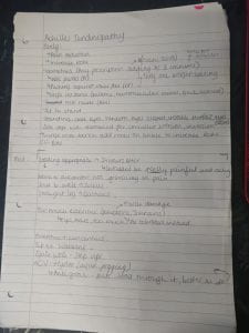
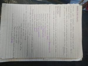
I spent one hour shadowing one of the masters students who had a disc prolapse. The therapist had seen the client before and so progress was measured first. The client had improved in SLR (10 degrees better), in ROMs, and in pain as well. They started with McKenzie’s extension pose and then moved on from there. Exercises were reviewed and they found them to be okay and still working at the moment. I enjoyed shadowing this session as the client was really into his rehabilitation plan. He understood everything and asked questions when needed. The remainder of the hours I spent revising for the upcoming exam which was based around a shoulder pathology and Achilles tendinopathy. I have added the sheets I created for the exam prep below, as seen the rotator cuff sheet is unfinished as I didn’t have time left in the clinic to finish it.

11/01/2019

Hours gained: 6

Total hours: 53

This week I only had two follow up appointments scheduled for me. The first appointment was spent massaging a lady who used to come in once a month for a deep massage into her back, shoulders, and neck. She worked a desk job and so always felt very stiff and tight. When testing ROM’s for her lumbar spine, shoulders, and neck I found that she was limited in all ranges. Before she came I made sure to read her notes and I realised that no one has ever given her any exercises to help with the tight muscles. During the STM we had a conversation about the effects of desk jobs and how it created muscle imbalances with her shoulders mainly. She asked questions and was interested in everything I had to say which showed that no one had told her before. After the STM, I gave her exercises which she could do at her desk as she mentioned that she didn’t have any time to do them. I gave her the Bruegger’s postural relief as I had been using that exercise whilst revising for the upcoming exam. Another suggestion I gave to her was to replace a chair with a swiss ball on some days or just for an hour every day. I also gave her side bends whilst seated, on the edge of the seat put your hands on the base of her back and try to push your elbows together. This exercise helped open up her chest as these muscles were very tight due to internally rotated shoulders.

Overall I think I did well with this client as I gave the exercises that were relevant and specific to her. No other therapist that had her gave her any exercises to help but I did. I also explained everything that was wrong to her with her posture and how you can use exercising and stretching to help that out. On the contrary, I could have also used posture tape to try and help with her internally rotated shoulders.

The other follow-up was a regular client who also came in once a month for his frozen shoulder. Last time I saw him his pain was improving so I expected it to either be the same or improve even more. However, when we went through the subjective assessment, he said that he was experiencing constant pain in his left anterior deltoid, bicep which occasionally went down to the elbow. He hadn’t done anything to aggravate it and hadn’t increased activity levels either. He felt the pain all through the day. The pain eased slightly after the first set of the exercises. In the objective assessment, I found that there was an increased pain when doing shoulder abduction from 60° to 120°. I started treatment with an STM on his left shoulder and biceps to help release the muscles a bit and relax them. He requested laser therapy as well so we did that next. We had another conversation about pain management and using either cold or heat to help him. When it came to the time of reviewing the exercises I could tell that the reps and sets were too high and so it was overworking his whole arm. I reduced the reps from 12 to 8-10, and the sets from 4 to 2-3. I gave him a range as I told him to see how he felt and then either do more or less. I also gave him a range as he comes in once a month and so I still wanted the exercises to work.

Overall I felt like I should’ve done more tests to see why the pain was going down to the elbow on some days. I should’ve added cervical myotomes just to check the nerves. I think I did well in realising that I overloaded him last time and so needed to take it a bit slower.

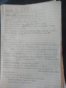
The other four hours were spent either revising, shadowing or helping with general clinic jobs. I shadowed two clients, one had lower back pain, and the other was suffering from tennis elbow. Lower back pain (LBP) was very common in the clinic as every 2/3 people will experience lower back pain in their lives (Hoy et al, 2010). During the first shadowing session, the therapist did an STM over the lower back and then reviewed exercises that had been given. He added weights to some exercises and took others out as she didn’t need them anymore. The other session included an STM over the forearm, tape for decompression, and then exercises that the client said he wasn’t going to do. I had a conversation with the therapist about giving him exercises and she said that she had done her job which was to prescribe them but she couldn’t force him to do them. I started going over my research and exam preparation for shoulder impingement (pictured below) and then helped with the general management of the clinic. I answered phones when the supervisor wasn’t there as well as folding fresh towels that came through.

Hoy, D., Brooks, P., Blyth, F. and Buchbinder, R. (2010) The Epidemiology of lower back pain. Best Practice & Research Clinical Rheumatology. Vol. 24, No. 6: 769-781.

14/12/2018

Hours gained: 6

Total hours: 47

I had one initial consultation appointment and two follow-up appointments this week. The first client, an initial consultation, came in complaining of pain in his right shoulder and neck. The pain came on about two weeks ago and was mainly felt on the top and outer side of the shoulder which when moving became a sharp pain into the arm. He mentioned he struggled with lifting his arm up over his head especially and that he can’t lie on that side when he’s sleeping. I asked what activities he did, he mentioned going to the gym every day for two hours mainly for resistance training. He had a desk job and so was sat in front of a computer from 9am-6pm. During the objective assessment, I found that the client struggled with abduction past 60°. He also didn’t like external rotation. The client had internally rotated and elevated shoulders. His ROM in his neck showed reduced range in left lateral flexion and rotation. The first pathology I wanted to clear was Thoracic Outlet Syndrome which I did through the special test. The differential diagnoses that I came up with were shoulder impingement or rotator cuff tear. I did the Hawkins-Kennedy test, Neers test, and internal rotation resistance strength test. The first two tests were positive and the last was negative. However, these tests are specific to shoulder impingement and also test positive for rotator cuff tears (Hegedus et al, 2008).

I also checked the scapular rhythm and found that there was scapular dyskinesis. The superomedial border of the scapular was also more superior. This led me to believe that due to scapular dyskinesis (type III) there’s an impingement of the shoulder. I started the treatment with an STM to the neck, shoulders, and thoracic back as they were very taut and he complained of “tight” muscles. I worked specifically into the upper trapezius and levator scapulae as they are commonly tight with this injury. I gave some stretches to help lengthen the levator scapulae and upper trapezius whilst giving strengthening and activation exercises for the lower trapezius. The stretches included seated upper trapezius stretch, seated levator scapulae stretch, and isometric ROMs of the neck for pain relief. The strengthening exercises included shoulder external rotation with theraband (both shoulders at the same time), Bruegger’s postural relief, and 90/90 scapular stabilisation. I also wanted to tape the scapula to help with the position however, we ran out of time and I had a client right after this one.

Overall I felt like I did really well in this session. The exercises I gave have high muscle activation and so were the best for the client. The 90/90 scapular stabilisation exercise was researched into by Maenhout et al (2016) and they found that exercises in prone had a higher muscle activation. The Brugger’s postural stretch helped in teaching the client the correct position his body should be in and was also very easy to incorporate into his life. During the STM I explained to the client that when training resistance, he needs to train his whole body and not focus on one area as he had been. I told him to start stretching the upper trapezius and strengthening the mid-lower fibres of it. I think the postural tape would have been beneficial for the client but due to poor time management, on my end, I couldn’t do it. I did add it into the future plan section though. I also felt like I should’ve checked cervical myotomes to clear cervical radiculopathy which could have been a differential diagnosis.

The next client I had was a follow-up and so I gave myself five minutes to read up on his notes to gain an understanding of what the pathology was and what the plan left behind was too. This case was a disc prolapse pathology and the treatment that was happening started off with an STM, then reviewed the exercises. When I went through the subjective assessment, the client mentioned that the STM helped him relax a little and that was what he mainly came here for. He mentioned that he doesn’t really do the exercises all the time and that they were a bit boring. I enquired about whether there was any pain, pins or needles down the legs and there wasn’t. I went through the objective assessment and found that ROMs and SLR were exactly the same as before and so there wasn’t much progression in that sense. However, the client did mention that his pain was reducing. I started the treatment with an STM and worked deep into the muscles as they were taut and adhesions were felt. I reviewed the exercises given to him to see that he didn’t know what they were as he never did them. He was given glute bridges and cobra position. I showed him how to do the exercises and then watched him do it. I added on another exercise to the program which was the cat and camel stretch. He laughed while he was doing it as it reminded him of twerking.

Overall I thought I could have added more imaginative exercises for him as he mentioned he found them boring. Hopefully, since he laughed at the cat and camel, he’d do it more often. I should’ve also started McKenzie’s extension position as I had used it on a lot of clients and it had worked.

I had this hour free and so I shadowed one of the other therapists. She had an initial consultation with an elderly gentleman, who was suffering from pain him his left ankle. He felt the pain on the lateral aspect of his ankle and walked with a cane. Through ROMs, we saw that there was an increase in inversion and a decrease in eversion. He mentioned that he used to fall over a lot and so they gave him a cane. She asked whether he used to go over his ankle a lot and he said that that was the cause of nearly all his falls. She did the chronic ankle instability questionnaire with him which showed he had chronic instability. She taped up his ankle to help provide some stability and then gave him strengthening exercises for his left leg. She started with seated calf raises and then worked a lot with balance and proprioception. She worked without the cane to try and help him strengthen and balance on that ankle. She started working on single-limb exercises and stretches straight away. I thought this was too soon as he found it hard to stand on two let alone one.

I had a follow-up appointment after this and found that it was one of my previous clients. I had treated him for tennis elbow the week before. After going through the subjective assessment, I found out that he had improved drastically. He had decreased the amount of golf he played found it had already started getting better. He also mentioned that the exercises he had were getting easier for him and he thought he needed harder ones. The objective assessment was very quick as I only needed to check his ROMs of wrist and elbow, and palpate the lateral epicondyle to see whether it was still as tender as before. The pain was still there at the end of the extension of the wrist and elbow however it was less. I massaged the forearm again and then used tape again for decompression. I reviewed the exercises and found that he didn’t need the resisted flexion and extension of the wrist anymore. I only added one more exercise as it had only been a week. I added weighted flexion and extension of the wrist. I started with very light weights and this seemed to be enough for him.

The other two hours were spent shadowing other therapists in the clinic. One case was a follow-up of medial tibial stress syndrome. In this case, he reviewed the exercises given and then increased the weight on farmers walks and added weights onto calf raises. The other case was also a follow-up but this client was suffering from facet joint stiffness in joints T5, T6, and T8. The client had a desk job and so was sitting down most of the time. He also wasn’t active at all and was overweight. The therapist tried to encourage the client to start getting active as it was putting too much pressure on his joints but he didn’t listen. This conversation helped me understand that if someone doesn’t want to do anything, then you can’t change their minds.

Hegedus, E. J., Goode, A., Campbell, S., Morin, A., Tamaddoni, M., Moorman III, C. T. and Cook, C. (2008) Physical examination tests of the shoulder: a systematic review with meta-analysis of individual tests.

Maenhout, A., Benzoor, M., Werin, M. and Cools, A. (2016) Scapular muscle activity in a variety of plyometric exercises. J Electromyogr Kinesiol. Vol. 27: 39–45.

07/12/2018

Hours gained: 7

Total hours: 41

This week I had a total of four clients, two initial consultations and two follow-up appointments. The first client I had was a female who was complaining of lower back pain which came on three days ago when she was picking up her son. My immediate thought after hearing this was that the injury was discogenic, due to the client being in a loaded flexed position. The pain was localised to the lumbar spine and hadn’t travelled down the leg or foot. The main objective findings were that the client had increased side flexion, to the left, in a standing position, and that the client’s pain increased when flexing but relieved a little when extending. I also tested straight leg raise (SLR) which was 70° on the right and 45° on the left. I explained to the client what I found and that it indicates the pathology to be disc prolapse. I also went on to explain the treatment and that extension exercises help promote the disc to go back into place. I used McKenzie’s treatment as I knew it had helped people I tried it on. Whilst she was in the position I massaged her lower back to help relieve the taut and spasming muscles. I gave her the cat and camel stretch, the cobra or leaning into the wall if she couldn’t do the cobra due to pain, knee hugs, and glute bridges. I also explained how she could continue the McKenzie treatment at home. I felt very confident and knowledgeable during the session as it was a spinal pathology which I feel comfortable with. I feel I could’ve improved the way I explained the d=injury as she was getting very worried and anxious about it. Next time I could use one of the spinal models in the clinic to help illustrate it. I could also help by not using complex words that may increase fear.

My next client was another initial consultation. The client came in with pain in their outer left elbow that he had for a couple of weeks. During the subjective assessment, the client mentioned that he doesn’t know how or when the pain started. His pain increased when lifting any object, especially his granddaughter who lives with him and when playing golf which he does four times a week. The client’s ROM was full in the wrist, elbow, and shoulder however his pain increased when he fulling flexed his elbow. During palpation, tenderness was expressed over the muscles in the forearm and the lateral epicondyle. I asked Alex to help me with this case as I hadn’t come across an elbow case yet and didn’t feel as comfortable in diagnosing and treating. After presenting the subjective and objective assessments to her, she said it’s a straightforward case of tennis elbow (lateral epicondylitis) due to the overuse of the tendons in the lateral aspect of the elbow. She told the client what the diagnosis was and also helped me with the treatment and discussed the need to reduce or stop how much golf done for a couple of weeks as it is increasing the pressure on the tendons. I started with an STM for the forearm then moved on to exercises; towel grips, towel twists, and resisted flexion and extension of the wrist. Kinesiology tape was also used to help decompress the forearm and elbow (Dilek et al., 2016). When the pain was too much the client was also advised to use a cold compress or ice pack to help alleviate some pain. During this session, I felt the complete opposite of my first client, I lacked confidence as well as knowledge. To improve this, I could try and revise injuries so I have at least a baseline of every injury. I should know how and what they present like. Another way to help me better myself would be to write differential diagnoses of different injuries and how they differ in presentation of symptoms or special tests.

I had the next hour free and so I researched into tennis elbow and what the main signs and symptoms are. Tennis elbow or lateral epicondylitis affects 3% of the population and occurs due to the overloading of the extensor tendons of the forearm (Bisset et al., 2011). Tennis elbow is a self-limiting injury which means that it can get better on its own, however, rest is required (Bisset et al., 2011). Alex mentioned that you’d usually find tennis elbow amongst people who either golf, are manual labourers, garden a lot, and playing musical instruments that involve repetitive movements of the wrist. Typical symptoms are; pain in fully extending either the wrist or elbow, weak grip strength, and pain when lifting and gripping (Tosti et al., 2013).

The third client this week was a follow-up. This was the third week I’ve seen this client for his frozen shoulder and bicep strain. The pain had reduced to more of a “niggling” pain. The ROM had increased but the pain was still felt through abduction of the shoulder, mainly 90° to 100°. There was also pain on external rotation. My treatment remained the same as I knew it was working. It started with an STM over the shoulder and biceps. Then I did laser therapy over the anterior and posterior head of the humerus and then I reviewed the exercises. This week the client thought that the exercise “throw salt over your shoulder” wasn’t helping or working anything so we decided to drop that one. I then added the original lateral raises as I had modified them for him before. This was also another way to see progression.

My next hour was free so I decided to shadow one of the other therapists to see how our treatments differ. The therapist had a case of lower back pain (LBP) and was an initial client. The therapist was struggling with diagnosing the injury which turned out to be facet joint dysfunction in L4 and L5. The therapist then did PAs on those levels to try and increase movement and then gave the client exercises.

I had a follow-up appointment after the shadowing session, who was suffering from facet joint stiffness in her cervical and thoracic spine. As per the plan I wrote last time, I first conducted central PAs on T4, T7, and T9 which I didn’t have time for during the last session. I did these movements in grade 2 for 60 oscillations for 3 sets which were also different from the last session. I increased the grade and the oscillations as the client could take it and it was the recommended amount (Ganesh et al., 2015). I also massaged the client’s upper back and shoulders to help work into the adhesions found last time. This client refused to take exercises last time so I knew I couldn’t do any more other than ask if she had changed her mind. She hadn’t. During this session, I felt like I had become confident again because I knew exactly what I had to do. It also helped that the client already felt better after one session.

The last hour was spent writing up notes, helping anyone that needed it and asking Alex about using the right words to communicate certain topics. I wanted to know how to talk to people that come in with discogenic problems as they often become very panicked and fearful that they’ll have to undergo surgery. I was told that it all comes down to educating them and helping them understand that its a very common pathology that only needs exercise to help it. This helped me a lot as I realised that as long as you reassure the client that they will be fine, they’ll listen to you. I also became more aware of how you can converse with a client in a more efficient way.

Bisset, L., Coombes, B. and Vicenzino, B. (2011) Tennis Elbow. British Medical Journal of Clinical Evidence. Vol. 6, No. 1: 1-35.

Dilek, B., Batmaz, I., Sariyildiz, M. A., Sahin, ., Ilter, L., Gulbahar, S., Cevik, R. and Nas, K. (2016) Kinesio taping in patients with lateral epicondylitis. Journal of Back and Musculoskeletal Rehabilitation. Vol. 29, No. 4: 853-858.

Ganesh, G. S., Mohanty, P., Pattnaik, M. and Mishra, C. (2015) Effectiveness of mobilization therapy and exercises in mechanical neck pain. An International Journal of Physical Therapy. Vol. 3, No. 2: 99-106.

Tost, R., Jennings, J. and Sewards, J.M. (2013) Lateral Epicondylitis of the Elbow. The American Journal of Medicine. Vol. 126, No. 4: 357.

30/11/2018

Hours gained: 3

Total hours: 34

This week I only had one client which was an initial consultation. This was the first hip client I had been given and so I was apprehensive as I know the hip is very complex. During the subjective assessment, the client mentioned that she felt pain over her left gluteus muscles (glutes) and the pain was felt when she was running, squatting and side lunging. I asked her to do those functional movements and I couldn’t find any fault with her technique at all. ROMs of her hips were all full, however, pain and was felt on the end range of abduction, internal rotation, and external rotation. I checked gluteus medius and minimus length and found the left side to be very limited when compared to the right. I also checked how long she could hold a glute bridge, the total time was 20 seconds. The Trendelenburg sign showed a dropped left hip which showed gluteus medius weakness. I gave the client some exercises to help strengthen the glute med as I knew that was causing pain and weakness. I gave the client clams, side abductions, mini wall squat (down to where she could feel discomfort but no pain) and resisted hip abduction. After researching these exercises I found that they weren’t the best exercises as they had the least muscle activation and so they didn’t work the gluteal muscles as well as I thought. The clam exercise only had 38% ± 18% MVIC (Maximum Voluntary Isometric Contraction) and the other exercises were among the high activation (Reiman et al., 2012).

I felt out of my comfort zone during that session and so I spent the other two hours revising other hip pathologies and how they present themselves. The first pathology I searched was Femoroacetabular Impingement (FAI). FAI is a when there’s extra growth either on the head of the femur and/or acetabulum. This causes the bones to rub against each other causing pain on movement and damage of the joint and surrounding structures (Clohisy et al., 2009). This could cause disability and early development of osteoarthritis (Clohisy et al., 2009). The symptoms of this injury include: pain felt in the anteromedial groin, limitations to daily living tasks, increased weakness in gluteal muscles, and gait abnormalities that may develop into a limp (Casartelli et al., 2011).

The next pathology I looked into was a groin strain. A groin strain is mainly caused by a strain in one of the adductor muscles (adductor longus, adductor magnus, adductor brevis, pectineus, and gracilis) that are medially situated to the thigh. The adductor muscles work together to adduct the hip, some of the muscles also help in other movements of the hip or knee. Strains have a sudden onset which is usually caused by high-speed exercise like; running, kicking, direction changes (Opar et al., 2012). The symptoms usually experienced with a groin strain are pain and tenderness in the groin region, pain when adducting the hip (especially at full ROM), pain when contracting adductors (thigh squeezes), and tightness or spasms in the groin area (Tak et al., 2016). These symptoms are very similar to that of a hamstring strain the only difference being where you feel the pain (Opar et al., 2012).

Casartelli, N. C., Maffiuletti, N. A., Item-Glatthorn, J. F., Staehli, S., Bizzini, M., Impellizzeri, F. M. and Leunig, M. (2011) Hip muscle weakness in patients with symptomatic femoroacetabular impingement. Osteoarthritis and Cartilage. Vol. 19, No. 7: 816-821.

Clohisy, J. C., Knaus, E. R., Hunt, D. M., Harris-Hayes. M. and Prather. H. (2009) Clinical Presentation of Patients with Symptomatic Anterior Hip Impingement. Clinical Orthopedics and Related Research. Vol. 467, No. 3: 638-644.

Opar, D. A., Williams, M. D. and Shield, A. J. (2012) Hamstring Strain Injuries: Factors that Lead to Injury and Re-Injury. Sports Medicine. Vol. 42, No. 3: 209-226.

Reiman, M. P., Bolgla, L. A. and Loudon, J. K. (2012) A literature review of studies evaluating gluteus maximus and gluteus medius activation during rehabilitation exercises. An International Journal of Physical Therapy. Vol. 28, No. 4: 257-268.

Tak, I., Glasgow, P., Langhout, R., Weir, A., Kerkhoffs, G. and Agricola, R. (2016) Hip Range of Motion id Lower in Professional Soccer Players With Hip and Groin Symptoms or Previous Injuries, Independent of Cam Deformities. The American Journal of Sports Medicine. Vol. 44, No. 3: 682-688.

22/11/2018

Hours gained: 5

Total hours: 31

This week consisted of two initial consultations and one follow-up. The first client was an initial consultation and so I had to wait to find out what injury I would be working on. The first client had upper back and neck pain that started a week ago. It was always present but felt more of a dull ache rather than pain itself. She tried heat and ice once but they didn’t help her. During the objective assessment, I found that her shoulders were internally rotated and that her shoulders were also elevated. All neck movements were not painful but they were limited. Thoracic movements created no pain however they were limited, overpressure movement was limited as well. During palpations, I found several adhesions along the rhomboids, trapezius, SCM, levator scapulae and erector spinae. These muscles also felt taut. I did PA’s on the neck and the thoracic spine which showed stiffness at C2, C4, C5, T4, T7 and T9 (all central). I concluded that the pain came from facet joint stiffness in the cervical spine and thoracic spine along with taut muscles and adhesions. The client mainly came in for an STM on the upper body and neck which is how I started my treatment. I also started PAs on the cervical spine. I tried to start my PA’s at grade two and for 60 oscillations, however, this was too much for the client. So, I did grade one PA, on C2 and C4, for four sets of 40 oscillations. I didn’t get time to continue PAs down to the thoracic spine and I thought it would have been too much anyway. The client wasn’t interested in any exercises and so prevented me from giving them to her.

During the first session, I realised I didn’t clear the shoulder or check shoulder movements which could have added more information if there were restricted movements. This could cause a problem as I might misdiagnose the client and disregard some differential diagnoses due to lack of information.

The next client was also an initial consultation. The client had two pathologies to look at; his neck and the tops of shoulders, and his left quadricep. Since he thought the neck and shoulders were a bigger problem we focused on that first. The neck pain came on 1o days ago when he was doing a forward roll and felt a sharp pain in his neck which later travelled to the shoulders. He always had some pain in his neck due to the very limited movement because of mechanical problems. In the objective assessment, I observed internally rotated and elevated shoulders and flexion in the thoracic spine. All movements of the neck increased pain and moved no more than 15 degrees, sometimes less. I made sure I cleared the shoulder this time as I know I didn’t in the previous client. There wasn’t any added pain and the ROM was also full. When palpating around the shoulders and neck, I found taut posterior deltoids, upper trapezius, and erector spinae. There was also stiffness in the facet joints, T4 to T10, when doing central PAs. I also checked the seat belt test which increased pain. I still hadn’t come to a conclusion of what it could be so I asked Alfie to come help me. Alfie came to the conclusion that it was a trapezius strain. He also told me to make sure I always ruled out thoracic outlet syndrome as it could have been a differential diagnosis. He also showed me the special test again. The client requested an STM due to being very “tight” so that was how I started my treatment. I gave resisted shoulder shrugs which is an isometric exercise said to reduce pain (Anwer and Alghadir, 2014; De Mey et al., 2009). I didn’t have time to do any PAs as part of the treatment so I added it to the future plan.

I had become better at focusing on one pathology at a time to help better manage my time and prevent rushing. However, during that session, I felt I had spent too much time on the objective assessment and should have called Alfie sooner to help me. This would’ve allowed for more treatment time. I spent the hour after this session researching and asking Alfie about how he knew it was specifically trapezius strain. I learned from Alfie that trapezius strains usually present themselves suddenly, the muscles are very tight and/or spasms and tight neck/shoulder complex. After researching a bit more, I found out that anxiety and low moods are also symptoms of trapezius strain/myalgia (Sjors et al., 2009). I struggled to find update research as well as research specifically about trapezius strain so I aimed my questions at Alfie instead. I wanted to know how to differentiate which muscle is strained. I was told that it’s based on movement and here the pain is. Palpating also helped differentiate the muscles too.

The follow-up client was a raiders player wanting to use the game ready and requested STM on his legs (quadriceps, hamstrings, and calves). I hadn’t used the game ready since the first year of university and so Alfie showed me how to use it again. Once that was set up, the client used it on his calves. The STM was done after as I didn’t want to warm the muscles up only to cool them down quickly again. I spoke about stretches to which he replied that he already did them and so I didn’t need to give him anymore. This was a very easy and quick appointment as I already knew I had done everything to help the client.

The last hour was spent shadowing another therapist. She had a client who had been coming for the past three weeks to use the anti-gravity treadmill. The client had a running injury which was caused by overtraining and had been that way for four weeks. The pain in the ankle and calf had reduced, ROM had increased and resisted movements had increased as well which showed signs of healing and strength as well. The therapist had the client running at 65% body weight at 11mph and on a slight incline to reduce the changes in gait.

Anwer, S. and Alghadir, A. (2014) Effect of Isometric Quadriceps Exercise on Muscle Strength, Pain, and Function in Patients with Knee Osteoarthritis: A Randomized Controlled Study. Journal of Physical Therapy Science. Vol. 26, No. 5: 745-748.

De Mey, K., Cagnie, B., Van De Velde, A., Danneels, L. and Cools, A. M. (2009) Trapezius Muscle Timing During Selected Shoulder Rehabilitation Exercises. Journal of Orthopaedic & Sports Physical Therapy. Vol. 39, No. 10: 743-752.

Sjors, A., Larsson, B, Dahlman, J., Falkmer, T. and Gerdle, B. (2009) Physiological responses to low-force work and psychosocial stress in women with chronic trapezius myalgia. BMC Musculoskeletal Disorders. Vol. 10, No. 1: 63.

15/11/2018

Hours gained: 5

Total hours: 26

This week I had two clients, both follow-ups. The first client came in with pain in her right shin which was felt during her training and walking. There was also a dull ache after training. This had been happening on and off for one year which was causing problems in her training sessions. The client trained four hours every day and had recently been in the Youth Olympics which had increased her pain. During the objective assessment, I tested ROM and resisted movements of the ankle and knee (they were all pain-free and all resisted movements were 5/5), palpated the ankle and calf (the pain was increased when palpating medial aspect of distal tibia 3/4 way down), recorded gait after running on the treadmill for five minutes and calculated cadence. The client said her pain began to travel further up the right tibia the longer she ran and the left shin began hurting too. When reviewing the footage, I found that she was a heel striker, her knees were varus due to fallen arches and her cadence was at 160 steps p/min. This told me that she put a lot of force through her ankle and knee joint through hitting her heel on the treadmill first (Daoud et al., 2012). Her varus came from the fact that she had fallen arches which misaligned her knees too. I came to the conclusion that she had Medial Tibial Stress Syndrome (shin splints) due to all the findings and symptoms. The treatment given to her was calf strengthening exercises. She was given; wall sits with heels raised, calf raises, single-leg glute bridges with the planted foot on the step, farmers walks with 1kg dumbbells, and arch contractions with toes planted. I ensured she understood how to do each exercise and corrected techniques when needed. We also had a conversation about training load as running and fencing were increasing the pain. Swimming seemed to help the client so suggested switching a few running or fencing sessions to swimming so she was still active but also allowed time to help the injury to heal. The plan for the next session is to get the client on the anti-gravity treadmill so she can run pain-free but still train as she usually would. This would also help her psychologically as she would feel as if she isn’t getting behind on training. I also want to help increase her cadence to no more than 170 steps p/min as this will help her decrease the amount of force she is putting through her body.

The next client came in with back pain and was a follow-up. Before I saw the client I took time in reading the notes left behind for him and found out that he had a disc prolapse at L4/L5. The previous treatment entailed STM on the lower back with some basic exercises as it was his first time. My plan for this session was to review ROM’s and the exercises. When the client came in, I ran through a subjective assessment where I learned that the pain was relatively the same. I went through the objective assessment and found that his ROM’s were exactly the same as the previous time. This was to be expected as the client had only been seen six days ago. I retested the straight leg raise, so I had that clinical marker, and saw a five-degree increase. I had recently gone through McKenzie exercises and procedures for lower disc problems with Gary and wanted to put that into practice. I explained what I was doing to the client and that I wanted him to stay in an extended position for at least ten minutes to help promote the disc back in place. I carried out STM on the client’s lower back whilst he was in this position as he requested it. I then gave him more exercises that were based around the extension of the lower back; cobra position, cat and camel, and leaning into the wall.

The remainder of my hours were built up in shadowing other students’ clients and learning more through what they say differently or how they conduct their objective assessments. During shadowing, one client came in with knee pain and the therapist carried a thorough subjective assessment and it came to my understanding that the client had medial meniscus damage. However, I don’t think the therapist thought that straight away. He continued to conduct a full objective assessment with special tests for everything and then asked me if I knew what it could be. I immediately said it was medial meniscus damage and we went from there.

This week I think I handled my sessions very well and I can see myself grow as a therapist. I already feel more confident with my diagnosis’s and when I’m not I know to ask for help. I really enjoyed the clients I had this week as I felt I put a lot of new techniques into training. I also learned a lot about the running world and common injuries that occur, through the first client and a conversation with Alex. I think in order to better myself I could become a lot clearer with my explanation of treatments and any tests conducted in the objective assessment. This is because it took me quite some time to explain what the McKenzie treatment was and what I wanted to do that session. Another thing I understood better this session was that everyone has different strengths and weaknesses and you can learn a lot from another person as they may excel at things I struggle with.

Daoud, A., Geissler, G. J., Wang, F., Saretsky, J., Daoud, Y. A. and Lieberman, D. E. (2012) Foot Strike and Injury Rates in Endurance Runners: A Retrospective Study. Medicine & Science in Sports & Exercise. Vol. 44, No. 8: 1325-1334.