Clinical Experience Tuesday 12th November 2019 – 5.5 hours (15:00-20:30): 103.5 total

Running total of hours: 103.5

Patient 1 – Patient Overview: regular weekly or fortnightly STM. Noticeable improvements in range of motion of neck and shoulders with a decrease in pain when putting on her coat. The progress in these sessions have been incredibly slow due to my inability to fully perform the treatment that may be most effective for the patient; mobilisations of the neck. This is due to the patient’s inability or discomfort in the thought of lying on the treatment couch and as such all STM is performed while the patient is sitting on the couch with legs over the side. The patient is happy with progress, knows the limitations in STM but reports positivity and provides good feedback for each session and wishes to continue the STM sessions on a weekly basis to supplement her physiotherapy appointments in between.

Patient 2 – Metatarsalgia follow up – No improvements but has recently developed new pain in lateral ankle, inferior to lateral malleolus over peroneal tendons. This has stopped the patient from running and fully engaging in rehabilitation and strengthening exercises.

According to findings by Männikkö & Sahlman (2017), orthotic inserts in the form of padding placed inside the shoes was effective as a treatment intervention to reduce pain and improve the scores of a questionnaire designed to evaluate lower limb pathology and has been reported as being one of the most commonly used foot scores for lower limb; The American Orthopaedic Foot & Ankle Society Forefoot Questionnaire (AOFAS). It must be noted however that although questionnaires in similar forms, such as the Self-Reported Foot and Ankle Score (SEFAS) and the Foot and Ankle Score (FAOS) have been found to be effective in correctly evaluating the presence of or change in dysfunction or injury presentation in the lower limb, the sheer range of foot disorders limits the ability to target certain pathologies which present with more specific symptoms. Although the AOFAS is a reliable measure, for future reference within a clinical setting, a questionnaire similar to the SEFAS may be a more useful tool as not only was it found to be a reliable and a feasible patient-reported outcome measure, it was also reported as being a much more user friendly method due to the time taken to complete; the AOFAS requires four comprehensive and resource demanding questionnaires, whereas the SEFAS requires scores on just pain, function and functional limitations but still maintained its reliability (Cöster et al., 2014).

Metatarsalgia can be the result of a number of risk factors, such as mechanical predisposed conditions such as pes planus, hammer toe, hallux valgus or hypermobility in the metatarsal joints and can be brought on by increased loading, fat pad alignment, but most interestingly rheumatoid arthritis (Männikkö & Sahlman, 2017). This patient is currently undergoing testing for Rheumatoid Arthritis after an appointment with his GP, due to a family history of the condition and the symptoms in his foot; it will be interesting to find out the results of this and how and if this may affect treatment. In the meantime, the use of padding or inserts are a good way to redistribute landing forces during walking or running and as such, this was given to the patient. A number of other studies including one by Besse (2016) have recommended the use of padding over the head of the metatarsal in order to create a new point in which the pressure is loaded upon and landing forces are dispersed over a larger surface area.
In order to fully test the effectiveness of this treatment, we did not change any other variables and kept his exercise program the same, as given at the previous session; if the patient reports a reduction in pain by the next session, then we can associate this with the padding and if there is no change, we can add another variable.

Figure 1 is of the typical location of metatarsalgia and is an accurate representation of where this patient felt most discomfort and pain and Figure 2 demonstrates when in the shoe the padding was placed.


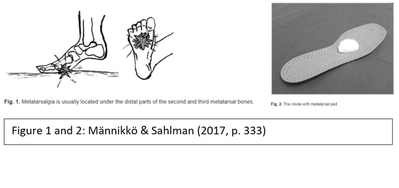
Although this method has been found to be of clinical use, it has also been found that a general insole with whole forefoot cushioning is effective in reducing and dispersing landing forces and peek pressure in the forefoot and is recommended over the more specifically located metatarsalgia padding as shown in Figure 2 (Hähni, Hirschmüller and Baur, 2016).
Because we did not have any orthotic inserts in the clinic to offer the patient, we constructed our own version of the padding shown in Figure 2.
If the patient reports some improvement with the extra padding, then we are able to better understand the mechanics of his injury. If there are no improvements, I can suggest the use of a more evenly constructed orthosis for our next option, such as the one used in a study by Hähni et al. (2016) as it eliminates patient application error and movement of the padding during activity; the location consistency may be compromised (Männikkö & Sahlman, 2017). The use of an orthosis may also provide enhanced positive effects in comparison to the padding, as shown by Hähni et al. (2016).

Patient 3 – STM treatment of the triceps surae group for the treatment of reduced dorsiflexion and pain in calf muscle.
This patient presented with significant feeling of “tightness” and associated pain of the triceps surae muscle group, more specifically the soleus (this was deduced through the isolation of the soleus muscle in a tandem stance lunge style stretch whereby the posterior leg being stretched is bent, eliminating the gastrocnemius muscle when the knee is flexed as this crossed over both the posterior knee onto the femur and the ankle via the calcaneal tendon).
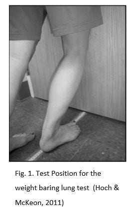
As mentioned in a previous post, the knee to wall test, also known as the lung test, is a useful measure for ankle dorsiflexion; it is a good indicator of muscle or joint restriction when compared with the contra lateral leg, or an indicator of improvements after treatments. It is commonly reported that tightness in the triceps surae group can cause a reduction in ankle dorsiflexion, which is a contributing factor in the future risk of injury (Hoch & McKeon, 2011).
When I performed this test in the clinic, I was not sure about the measurements that were of significance. According to Hoch & McKeon (2011), asymmetry can be determined by a difference of >1.5cm between each ankle. In this case, asymmetry was not present as the patient presented with bilateral tightness and subsequent restrictions in ankle dorsiflexion in both ankles.
In order to help reduce risk of future ankle injury and reduce pain and tightness, I administered treatment to lengthen the triceps surae muscles by way of soft tissue massage, stretching, exercises and derived a take home plan to maintain these treatment effects, which also involved self-administered soft tissue massage such as foam rolling (Wiewelhove et al., 2019).
Soft tissue therapy is widely used in the treatment of soft tissue injuries, but the efficacy of its use is also often disputed. There is a great body of evidence to disregard the effectiveness of this modality, however conversely there are also findings proving its effectiveness; the use of soft tissue massage should therefore be advocated only on a case by case basis and measures to test it’s effectiveness put in place.
According to findings by Thomson, Gupta, Arundell and Crosbie (2015), deep soft tissue massage is not an effective treatment for muscle stiffness or restricted ankle dorsiflexion; no significant differences in extensibility or reduced stiffness was found. This study was conducted using a reportedly highly reliable test method but only included 29 healthy subjects without any musculoskeletal injury. Although this study found massage as ineffective on a small sample of healthy subjects, these findings may not be representative of the wider population and research needs to be done on the effects on injured or dysfunctional tissue. Early research by McKechnie, Young and Behm (2007) on the effectiveness of two different massage techniques found that just three minutes of massage by tapotement or petrissage over the muscles that plantarflex the ankle (peroneal longus and brevis, gastrocnemius and soleus) improved ankle flexibility, but not with an associated reduction in power, justifying the use of this modality for at least a short term treatment option. Interestingly, no differences were found between the two types of massage, therefore an application for either can be effective.
In a study on 50 female hockey players, dynamic soft tissue massage was compared to regular massage in the treatment of increasing hamstring length with both control and intervention group experiencing increases after treatment, suggesting the benefits of massage in general on increasing tissue length in muscles (Hopper et al., 2005).

More recent research on a larger sample size of 60 subjects by Stefansson, Brandsson, Langberg and Arnason (2019) for example, found that pressure point massage was effective in the treatment of Achillies Tendinopathy, an injury presenting with muscular tightness in the triceps surae group as a risk factor. The positive effects of pressure massage, therefore could imply that mechanical effects do occur as a result of STM, as previously refuted by Thomson et al. (2015).
Although there is conflicting evidence and varying recommendations on the effectiveness and efficacy of STM for exercise recovery, ROM, exercise performance and stiffness, the use of STM for the reduction of pain has been reported and evidenced in studies such as a study by Romanowski et al. (2012), showing that massage, both therapeutic and deep tissue can have positive effects on pain in individuals with chronic lower back pain and Kumar, Beaton and Hughes (2013) who found that massage produced short term pain reduction in chronic lower back pain.

Interestingly, in a review on the effects of STM by Piper et al. (2016), soft tissue therapy was found to be effective in some injuries, such as lateral epicondylitis and plantar heel pain, however it was highlighted that movement education was also effective in treating lateral epicondylitis, implying neuromuscular adaptations. This posed the question of whether any reported effects of massage is correlated to the physiological changes in the soft tissue, or the neuromuscular interpretation of muscular length, with reports published on the effects on neurological effects of massage (Sefton, Yarar, Carpenter and Berry, 2011).
Other factors in the reduction of pain can be discussed, such as the combination of physiological and psychological effects.
It has been reported that massage can have a variety of other benefits as well as the reduction of pain, including improvements in stress levels, emotional wellbeing and physical recovery; found in a study by Adams, White and Beckett (2010) when administered in the early stages of their treatment or care in a hospital setting.

Figure 1 shows the test procedure of the test used as an objective marker (Hoch & McKeon, 2011). Not only was I able to confirm the physiological adaptations and effectiveness of the massage treatment by way of qualitative feedback of reduced pain from the patient, but the patient was able to see for herself the effectiveness of the session, hopefully providing her with motivation to maintain a home program outside of the clinic setting.
I will ask the patient to perform the test again in her next session, as this may be a suitable indicator of exercise and stretching adherence over the coming weeks.

Patient 4 – STM of lower back
Patient Overview – This patient is very physically active, attending the gym most days. He presented with chronic lower back pain which is being managed and treated by a variety of care providers including the sports therapy clinic at Marjon. The patient has already been assessed and asked specifically for a soft tissue massage only as a means to compliment the stretching and exercise program he was prescribed at his previous treatment sessions.

Non-specific Low Back Pain (NSLBP) has a prevalence of up to 33% and accounts for 85-90% of lower back pain (Kachanathu et al. (2014). Although most individuals with lower back pain respond well to treatment in the early stages, according to Kachanathu et al. (2014) up to 15% end up with a chronic condition.
As the patient has felt improvements and positive effects of soft tissue massage for his lower back, he was satisfied with the nature of the treatment. I made sure to ask for feedback throughout the treatment, ensuring that I was applying the desired pressure.
I checked the patients ROM and updated his records to reflect this so that improvements can be made and used pain and patient reported stiffness as marker for improvement and performed deep tissue massage and myofascial release to his whole lower back and thoracic spine as well as deep tissue massage through his glutes and external rotators. Deep tissue massage has been reported as more effective than therapeutic massage in the treatment of lower back pain, so I needed to ensure I was applying the appropriate pressure. I mobilised his lower spine using posterior anterior grade IV oscillations to help reduce pain and increase ROM in lumbar extension and flexion, as found effective in the treatment of NSLBP (Shah & Kage, 2016).
In order to help the patient to improve his LBP on a more long term time scale, mobility exercises such as the cat camel, knee rolls, knee hugs and strengthening of the abdominal and gluteus muscles were given and stretching of the hip flexors, hamstrings and back were advised as recommended by a study finding the positive outcome of this treatment in patients with chronic LBP (Kachanathu et al., 2014).

Extra hour between patients – I spend this hour trying to increase my dorsiflexion range of motion through a range of interventions; mobilisations with movement using a resistance band and by performing squats. I practiced performing the knee to wall test and found noticeable improvements using both methods. I am aware of my dorsiflexion restriction, particularly in my right ankle due to previous lateral ankle sprain injuries and so I knew that joint mobilisations would be more effective than general soft tissue massage of the trceps surae group alone.
From this practice, I was able to fully appreciate the need for the non tested foot to have the heel flat on the floor so as to no wrongly add further range to the tested ankle. Trying out these tests and exercises myself is a good way to understand them in a better way in order for me to demonstrate them better and more efficiently to my patients in future.

References –

Adams, R., White, B., & Beckett, C. (2010). The effects of massage therapy on pain management in the acute care setting. International Journal of Therapeutic Massage and Bodywork: Research, Education, and Practice, 3(1), 4–11. https://doi.org/10.3822/ijtmb.v3i1.54

Besse, J.-L. (2016). Review article Metatarsalgia. 103, 29–39. https://doi.org/10.1016/j.otsr.2016.06.020

Cöster, M. C., Bremander, A., Rosengren, B. E., Magnusson, H., Carlsson, Å., & Karlsson, M. K. (2014). Validity, reliability, and responsiveness of the Self-reported Foot and Ankle Score (SEFAS) in forefoot, hindfoot, and ankle disorders. Acta Orthopaedica. https://doi.org/10.3109/17453674.2014.889979

Hähni, M., Hirschmüller, A., & Baur, H. (2016). The effect of foot orthoses with forefoot cushioning or metatarsal pad on forefoot peak plantar pressure in running. Journal of Foot and Ankle Research, 9(1), 7–13. https://doi.org/10.1186/s13047-016-0176-z

Hoch, M. C., & McKeon, P. O. (2011). Normative range of weight-bearing lunge test performance asymmetry in healthy adults. Manual Therapy, 16(5), 516–519. https://doi.org/10.1016/j.math.2011.02.012

Hopper, D., Conneely, M., Chromiak, F., Canini, E., Berggren, J., & Briffa, K. (2005). Evaluation of the effect of two massage techniques on hamstring muscle length in competitive female hockey players. Physical Therapy in Sport, 6(3), 137–145. https://doi.org/10.1016/j.ptsp.2005.04.003

Kachanathu, S. J., Alenazi, A. M., Seif, H. E., Hafez, A. R., & Alroumim, A. M. (2014). Comparison between Kinesio taping and a traditional physical therapy program in treatment of nonspecific low back pain. Journal of Physical Therapy Science, 26(8), 1185–1188. https://doi.org/10.1589/jpts.26.1185

Kumar, S., Beaton, K., & Hughes, T. (2013). The effectiveness of massage therapy for the treatment of nonspecific low back pain: A systematic review of systematic reviews. International Journal of General Medicine. https://doi.org/10.2147/IJGM.S50243

Männikkö, K., & Sahlman, J. (2017). The Effect of Metatarsal Padding on Pain and Functional Ability in Metatarsalgia. Scandinavian Journal of Surgery. https://doi.org/10.1177/1457496916683090

McKechnie, G. J. B., Young, W. B., & Behm, D. G. (2007). Acute effects of two massage techniques on ankle joint flexibility and power of the plantar flexors. Journal of Sports Science and Medicine, 6(4), 498–504.

Piper, S., Shearer, H. M., Côté, P., Wong, J. J., Yu, H., Varatharajan, S., … Taylor-Vaisey, A. L. (2016). The effectiveness of soft-tissue therapy for the management of musculoskeletal disorders and injuries of the upper and lower extremities: A systematic review by the Ontario Protocol for Traffic Injury management (OPTIMa) collaboration. Manual Therapy, 21, 18–34. https://doi.org/10.1016/j.math.2015.08.011

Sefton, J. E. M., Yarar, C., Carpenter, D. M., & Berry, J. W. (2011). Physiological and clinical changes after therapeutic massage of the neck and shoulders. Manual Therapy. https://doi.org/10.1016/j.math.2011.04.002

Shah, S. G., & Kage, V. (2016). Effect of seven sessions of posterior-to-anterior spinal mobilisation versus prone press-ups in non-specific low back pain-randomized clinical trial. Journal of Clinical and Diagnostic Research, 10(3), 10–13. https://doi.org/10.7860/JCDR/2016/15898.7485

Stefansson, S. H., Brandsson, S., Langberg, H., & Arnason, A. (2019). Using Pressure Massage for Achilles Tendinopathy: A Single-Blind, Randomized Controlled Trial Comparing a Novel Treatment Versus an Eccentric Exercise Protocol. Orthopaedic Journal of Sports Medicine, 7(3), 1–10. https://doi.org/10.1177/2325967119834284

Thomson, D., Gupta, A., Arundell, J., & Crosbie, J. (2015). Deep soft-tissue massage applied to healthy calf muscle has no effect on passive mechanical properties: A randomized, single-blind, cross-over study. BMC Sports Science, Medicine and Rehabilitation, 7(1), 1–8. https://doi.org/10.1186/s13102-015-0015-8

Wiewelhove, T., Döweling, A., Schneider, C., Hottenrott, L., Meyer, T., Kellmann, M., … Ferrauti, A. (2019). A meta-analysis of the effects of foam rolling on performance and recovery. Frontiers in Physiology. https://doi.org/10.3389/fphys.2019.00376


Clinic Experience Monday 11th November 2019 – 6 hours (15:00-21:00): 98 total

Running total of hours: 98

Patient 1 – Patient Overview: This patient is a student therapist and presented with acute and severe back pain and neurological symptoms of sciatic down his left side. Sudden Onset 2 hours after volleyball training after sitting down. Very restricted in all ROM and experiencing 9/10 levels of pain.

Neurodynamic are still a weak area of my knowledge and understanding and so the practical application of testing and treatment is a real challenge for me within my practice. I am confident in the methods used to test for lower back neurological dysfunction but I am not able to interpret these findings or follow through an effective, thorough assessment that boasts accuracy or specificity.
My supervisor helped to work through the assessments for root nerve impingements, but even when observing, I was unable to fully appreciate the mechanisms used and outcomes observed. I also find treatment in response to findings very challenging.

The Straight Leg Raise is performed as demonstrated by the Physio tutors:

https://www.youtube.com/watch?v=LdAD9GNv8FI

According to a review by Capra et al. (2011) the straight leg raise was not found to be an effective in identifying lumbar disc herniations with a sensitivity of 0.36 and a specificity of 0.74.
Two other commonly used neurodynamic tests for lower limb pathological neuropathy are as follows and are useful tools in the assessment of patients with symptoms with radicular leg pain indicating nerve root impingement or lumbar disc herniation.
The Slump Test as shown in the following video by the Physiotutors, has been shown to have a sensitivity of 0.91 and a specificity of 0.70 (Urban & Macneil, 2015):

https://www.youtube.com/watch?v=HFGfP84uwEo

And the Prone Knee Bend as demonstrated in this video by the Physiotutors.

https://www.youtube.com/watch?v=4VxKyPRq6HA

These two tests alone have reasonable sensitivity and specificity, however in research conducted in a pilot study by Trainor & Pinnington (2011), a technique known as the slump knee bend test as shown in figure 1 is a variation on the prone knee test and found to have a sensitivity of 1.00 and specificity of 0.83 at identifying lumbar root compression at L4. Although it was noted that this test may not be able to identify compression at other lumbar locations, it is recommended as a slightly more reliable test for the presence of neurological pathology and as such could be used in conjunction with other neurodynamic tests. The test was carried out as shown in Figure 1.


From these tests we found positive signs and indicators of nerve root impingement, but I was unsure as to how to treat my patient based on these findings. Because of the acute nature of the injury, the likelihood of muscle guarding and spasms and the intensity of pain, it would have been difficult to fully assess this patient at this session. I did, however, apply STM to his lower back to help relieve tension from the surrounding soft tissue and to help relieve the pain. The patient was advised to return to the clinic for some traction to decompress the spine in an attempt to reduce pain and improve function in the lumbar spine with research suggesting that traction even in the form of the straight leg raise with added traction is an effective treatment modality, especially when combined with other treatments such as home based strengthening exercise programs (Cho, Lee and Hwangbo, 2015; Gǔlşen, Atici, Aytar and Sahin, 2018; Meszaros et al., 2000; Pawar & Metgud, 2014). It is useful to know that in the absence of a traction machine or if the patient was of large body size, posterior to anterior mobilisations have also been found as equally effective as traction with straight leg raise. Another pain relief option that I did not consider but are recommended could be the use of a TENS machines (Pawar & Metgud, 2014).
Fortunately, however this clinic has a mechanical traction machine which is readily available and as such proved very useful in this particular case with this treatment being found to significantly change pain levels and function in patients with nerve root compression when 12 treatments were performed over 6 weeks.

According to NICE Guidelines, as summarised by Bernstein Malik, Carville and Ward (2017), manual therapy and exercise programs were advised as well as other modalities such as stretching and mobilisations, all of which I will hope to incorporate into a rehabilitation program for the patient as the acute nature of the injury subsides.

Patient 2 – This patient presented with lateral knee pain with the clinical subjective notes as follows;
Started running in January 11/24 ago, couch to 5km. increasing mileage and groups. Started getting “niggles” in ankle, no niggles since 3/12 ago.
Off road event, slipped and now sharp pain in knee. 5 days of Px now fine. Has had week off running.
Was recommended a sports massage. Booked 2/52 ago.
10km, training for 1/2 marathon. Increasing running, 11.5miles.
Currently 25miles/week.
1/52 ago Px in lat. R knee. Px in morning, eases through day, Px again in evening.
Running stabbing Px 7/10, had to stop running. Now no Px.
Stairs Px going down stairs.
Px when injury at worse. OK now.

Although this patient had requested a sports massage, it became apparent early on in the subjective assessment that this modality alone would not prove the most effective in this case and a more comprehensive assessment was required. As the patient was unsure as to the nature of a sports massage and visited the clinic with the clear objective of an injury diagnosis or treatment, I continued to perform a full assessment instead.
From an objective assessment, I noted that the patient had significant pes planus in the same foot as his knee pain. Pes planus is a common foot deformity also known as flat feet and is commonly caused by weakness in the surrounding muscular, such as the plantar and dorsiflexors, inverters, and evertors, but more specifically the tibialis posterior muscles, which, when weakened can present with a condition recognised as posterior tibial tendon dysfunction (Erol et al., 2015).

According to a findings from a study by Lee & Choi (2016) and Ashford, Mathieson and Rom (2016) strengthening exercises of the foot intrinsic muscles and tibialis posterior were effective in treating possible causes or symptoms of pes planus by way of reducing foot stability, plantar pressure and balance with an administered rehabilitation program of six weeks included five 30minute sessions. It has also been reported in a study on runners in particular that strengthening of the intrinsic and extrinsic foot muscles is an effective treatment modality for individuals with pes planus who present with associated overuse injuries (Ridge et al., 2018).

During the session I attempted to demonstrate an exercise to help strengthen the patient’s arches but failed terribly; the movements of arch squeezes are subtle and difficult to explain and as such I did not feel as though the patient was confident in his understanding of the exercises.
I have since been able to find some more exciting exercises to use for patients to do and hopefully with a little more enjoyment too, which may also help to increase exercise adherence.
Exercises such as picking up objects with feet, folding clothing or towels with feet and walking on the lateral side of feet were suggested by Erol et al. (2015) as well as the more-simple resistance band exercises and toe heel walking.

Shadowing Ankle Taping 
In between patients, I decided to shadow another therapist who was treating a patient as part of his placement with the Marjon University Netball team. This patient had suffered a mild ankle sprain two weeks prior to this session and was hoping to return to training today, but with appropriate level of participation. In order to take part, additional support by means of rigid ankle taping was applied, the method used was that of the figure of 8 shown in the video below and as used in a study by Halim-Keranegara, Raymond, Hiller, Kilbreath and Refshauge (2017) and in figure 1.

https://www.youtube.com/watch?v=16lF6hOB4bs

Although the effects of ankle taping by way of mechanical support has been widely disputed, with studies suggesting it as beneficial such as Kuni, Mussler, Kalkum, Schmitt and Wolf (2016) and those that have found it is not, such as Jaffar et al. (2016), there are some other benefits beyond these physical effects. Psychological elements, including an athlete’s perceived ability to perform and self-efficacy were reportedly improved after mechanical taping was applied and as such the use of tape may be useful in aiding a quicker return to sport (Halim-Kertanegara et al., 2017).

The therapist used non elastic rigid taping for this procedure but as reported by Abián-Vicén, Alegre, Fernández-Rodŕiguez and Aguado (2009), with the same supporting effects, elastic taping has been found to be more comfortable and less restricting but without compromising the mechanical effects of the tape. The patient was quick to complain about the discomfort, not in her ankle, but by the feeling of the tape, so this study was able to provide evidence for the use of a more comfortable alternative. In future, if mechanical support is my objective then this taping may be slightly more comfortable.
It is thought that kinesiology tape is an effective treatment to help improve functional performance in the ankle, help improve balance and proprioception and help prevent lateral ankle sprains (Jackson, Simon and Docherty, 2016; Lee & Lee, 2016; Y. Wang et al., 2018). One of the physiological effects of the tape is the neurological responses from the mechanoreceptors in the ankle with tape; the feeling of the tape on the ankle becoming a more efficient stimulus for quicker muscle reaction times of the peroneals in particular and in individuals with chronic ankle instability, either caused by or which has caused muscle weaknesses in the ankle. Research by Jaffar and Li (2016) for example, found greater increases in muscle activation of the peroneal longus muscle during dynamic movements in subjects with functional ankle instability with the presence of rigid tape compared with subjects with kinesiology tape or no tape.

Patient 3 – This patient presented with Lower back and with a prevalence of up to 33% in the general population (Kachanathu, Alenazi, Seif, Hafez and Alroumim, 2014), this patient is one of many who I come across for soft tissue therapy treatment in the clinic. I treated this patient with evidence based deep tissue massage and mobilisations as well as prescribing a suitable exercise and stretching program to help strengthen and maintain more pain free ROM (Kachanathu et al., 2014; Shah & Kage, 2016). However, what I found most interesting in this case was the request for traction of the hip by way of unilateral pulling of each leg at the ankle. It has been found that treatment aimed at the hip joints are effective in the management of chronic lower back pain (Burns, Mintken, Austin and Cleland, 2011). This patient self-reported improvements in his symptoms after this treatment. I have performed traction type mobilisations to the SI Joint at my external placement and have developed an effective technique for this, but this was my first experience of traction of the hip.
In order to perform this, I grasped the patient’s ankle and pulled, producing and longitudinal distraction of the hip and repeated this on both legs. This method has been suggested for patients who require a general stretch of the hip joint capsule (Reiman & Matheson, 2013). Figure 2 is a version of this distraction technique that can be performed at home so the patient can continue with this treatment at home to maintain progress and reduced levels of pain in between the treatment sessions.

Patient 4 – This patient was a follow up from the previous week (Patient 3, Tuesday 5th November). Nothing has changed from the previous week of treatment for possible achillies tendinopathy but as we ran out of time for STM, she had returned to receive this. As I forgot to test any objective measures, before I performed treatment as used in Stefansson, Brandsson, Langberg and Arnason (2019), I made sure to carry out the knee to wall test, which showed bilateral shortening in both legs and a significant inability to gain a reasonable distance from her foot to the wall, indicating the need to increase either joint mobility or probably more likely, the presence of gastrocnemius and soleus dysfunction by way of reduced length or flexibility. There is an abundance of research on the methods of achillies tendon rehabilitation, however they are often conflicting and are continuing to develop. Sports massage treatments and programs including eccentric and/or concentric exercises have been studied and reviewed, with eccentric exercises most often the more preferred option. Research conducted by Malliaras et al. (2013), for example found that no one method is more effective than the other but suggested eccentric-concentric as a foundation of treatment. This study, along with many other, highlight the need for further research into the mechanical effects of various loading programs; a possible reason why treatment methods remain undecided. A more recent study by Chaudhry, Morrissey, Woledge, Bader and Screen (2015) found that eccentric exercises were more effective than concentric exercises of the triceps surae muscle group in the rehabilitation of achillies tendinopathy but other studies within that same year found either method to be equally as effective with no significant differences between subject groups (Beyer et al., 2015). A single-blind randomised control study on the effects of massage on achillies tendinopathy by Stefansson et al. (2019) reported that a commonly accepted risk factor for achillies overuse injuries is muscular tightness in the triceps surae group and associated reduction in ankle ROM. Furthermore, although eccentric strengthening is widely reported as the most effective treatment for this injury, this study found that pressure massage was as effective as eccentric exercises and potentially allowed for a quicker resolution in symptoms, as this modality can result in earlier and more immediate effects. Interestingly, however, this research did not find reason to combine the two treatments as further effects of this were not significantly increased.

In order to maintain the effects of the STM at home, I suggested that the patient use a foam roller at home on a daily basis, as this has been found to be effective in reducing muscle pain and increasing flexibility (Wiewelhove et al., 2019).
This session was also a good opportunity to ensure that the patient was performing the prescribed exercises with the correct routine, having had a week to get to grips with the program and discover any areas that may not have been fully understood. Often, patients rebook appointments for weeks ahead and as such, any issues with the prescribed programs cannot be addressed early enough. In this case, the patient queried the side lying abductor exercise (prescribed for suspected weakness in the hip abductors) as she was unsure as to whether her hips should be pushed forward or allowed to drop back; I informed her that in order to isolate the Gluteus Medius muscle, the hips must be pushed forward, as opposed to relaxing the hip back, which activates the Tensor Fascia Latae muscle. In order to ensure that the correct muscles are being used, I instruct the patient to place the tips of their fingers over their Gluteus Medius muscle so that they can feel it contracting during the movement.

Spare time between patients – as neurodynamic testing is a significant area of weakness for me, both in knowledge and practice, as well as the researching behind these, I was able to practice with a partner as fortunately the clinic with busy but with some cancelled appointments. Talking through procedures and methods with other students always seems to take longer than when we are shown directly from the clinic manager or from a lecturer, however by trying to figure out how to do something ourselves, we learn all of the way not to do it and have to understand the practice better in order to understand how it works. I find this time between patients incredibly useful and if there are other students around to help, they are always able to add their own strengths to our practice and they are always willing to help.

References –

Abián-Vicén, J., Alegre, L. M., Fernández-Rodŕiguez, J. M., & Aguado, X. (2009). Prophylactic ankle taping: Elastic versus inelastic taping. Foot and Ankle International. https://doi.org/10.3113/FAI.2009.0218

Ashford, R., Mathieson, I., & Rome, K. (2016). Conservative Interventions for mobile Pes Planus in Adults: a systematic review. Revista Internacional de Ciencias Podológicas. https://doi.org/10.5209/rev_ricp.2016.v10.n2.52304

Bernstein, I. A., Malik, Q., Carville, S., & Ward, S. (2017). Low back pain and sciatica: Summary of NICE guidance. BMJ (Online). https://doi.org/10.1136/bmj.i6748

Beyer, R., Kongsgaard, M., Hougs Kjær, B., Øhlenschlæger, T., Kjær, M., & Magnusson, S. P. (2015). Heavy slow resistance versus eccentric training as treatment for achilles tendinopathy: A randomized controlled trial. American Journal of Sports Medicine, 43(7), 1704–1711. https://doi.org/10.1177/0363546515584760

Burns, S. A., Mintken, P. E., Austin, G. P., & Cleland, J. (2011). Short-term response of hip mobilizations and exercise in individuals with chronic low back pain: A case series. Journal of Manual and Manipulative Therapy. https://doi.org/10.1179/2042618610Y.0000000007

Capra, F., Vanti, C., Donati, R., Tombetti, S., O’Reilly, C., & Pillastrini, P. (2011). Validity of the straight-leg raise test for patients with sciatic pain with or without lumbar pain using magnetic resonance imaging results as a reference standard. Journal of Manipulative and Physiological Therapeutics. https://doi.org/10.1016/j.jmpt.2011.04.010

Chaudhry, S., Morrissey, D., Woledge, R. C., Bader, D. L., & Screen, H. R. C. (2015). Eccentric and concentric exercise of the triceps surae: An in vivo study of dynamic muscle and tendon biomechanical parameters. Journal of Applied Biomechanics. https://doi.org/10.1123/JAB.2013-0284

Choi, J., Lee, S., & Hwangbo, G. (2015). Influences of spinal decompression therapy and general traction therapy on the pain, disability, and straight leg raising of patients with intervertebral disc herniation. Journal of Physical Therapy Science. https://doi.org/10.1589/jpts.27.481

Erol, K., Karahan, A. Y., Kerimoğlu, Ü., Ordahan, B., Tekin, L., Şahin, M., & Kaydok, E. (2015). An important cause of pes planus: the posterior tibial tendon dysfunction. Clinics and Practice. https://doi.org/10.4081/cp.2015.699

Gǔlşen, M., Atici, E., Aytar, A., & Sahin, F. N. (2018). Effects of traction therapy in addition to conventional physiotherapy modalities on pain and functionality in patients with lumbar disc herniation: Randomized controlled study. Acta Medica Mediterranea. https://doi.org/10.19193/0393-6384_2018_6_315

Halim-Kertanegara, S., Raymond, J., Hiller, C. E., Kilbreath, S. L., & Refshauge, K. M. (2017). The effect of ankle taping on functional performance in participants with functional ankle instability. Physical Therapy in Sport, 23, 162–167. https://doi.org/10.1016/j.ptsp.2016.03.005

Jackson, K., Simon, J. E., & Docherty, C. L. (2016). Extended use of kinesiology tape and balance in participants with chronic ankle instability. Journal of Athletic Training, 51(1), 16–21. https://doi.org/10.4085/1062-6050-51.2.03

Jaffar, M. R., Jaafar, Z., & Li, G. S. (2016). Peroneus longus activity in different types of taping: athletes with ankle instability. Revista Brasileira de Medicina Do Esporte. https://doi.org/10.1590/1517-869220162203142486

Kachanathu, S. J., Alenazi, A. M., Seif, H. E., Hafez, A. R., & Alroumim, A. M. (2014). Comparison between Kinesio taping and a traditional physical therapy program in treatment of nonspecific low back pain. Journal of Physical Therapy Science, 26(8), 1185–1188. https://doi.org/10.1589/jpts.26.1185

Kuni, B., Mussler, J., Kalkum, E., Schmitt, H., & Wolf, S. I. (2016). Effect of kinesiotaping, non-elastic taping and bracing on segmental foot kinematics during drop landing in healthy subjects and subjects with chronic ankle instability. Physiotherapy (United Kingdom). https://doi.org/10.1016/j.physio.2015.07.004

Lee, D., & Choi, J. (2016). The Effects of Foot Intrinsic Muscle and Tibialis Posterior Strengthening Exercise on Plantar Pressure and Dynamic Balance in Adults Flexible Pes Planus. Physical Therapy Korea. https://doi.org/10.12674/ptk.2016.23.4.027

Lee, S. M., & Lee, J. H. (2016). Effects of ankle eversion taping using kinesiology tape in a patient with ankle inversion sprain. Journal of Physical Therapy Science. https://doi.org/10.1589/jpts.28.708

Malliaras, P., Barton, C. J., Reeves, N. D., & Langberg, H. (2013). Achilles and patellar tendinopathy loading programmes: A systematic review comparing clinical outcomes and identifying potential mechanisms for effectiveness. Sports Medicine. https://doi.org/10.1007/s40279-013-0019-z

Meszaros, T. F., Olson, R., Kulig, K., Creighton, D., & Czarnecki, E. (2000). Effect of 10%, 30%, and 60% body weight traction on the straight leg raise test of symptomatic patients with low back pain. Journal of Orthopaedic and Sports Physical Therapy. https://doi.org/10.2519/jospt.2000.30.10.595

Pawar, A. H., & Metgud, S. (2014). “ Comparative Effectiveness of Mulligan ’ s Traction Straight Leg Raise and Bent Leg Raise in Low Back Ache with Radiculopathy ” – A Randomized Clinical. International Journal of Science and Research.

Reiman, M. P., & Matheson, J. W. (2013). Restricted hip mobility: clinical suggestions for self-mobilization and muscle re-education. International Journal of Sports Physical Therapy.

Ridge, S., Henderson, A., Bruening, D., Jurgensmeier, K., Olsen, M., Griffin, D., … Davis, I. (2018). Midfoot Angle Changes During Running After an 8-week Foot Strengthening Program. Foot & Ankle Orthopaedics, 3(3), 2473011418S0040. https://doi.org/10.1177/2473011418s00405

Shah, S. G., & Kage, V. (2016). Effect of seven sessions of posterior-to-anterior spinal mobilisation versus prone press-ups in non-specific low back pain-randomized clinical trial. Journal of Clinical and Diagnostic Research, 10(3), 10–13. https://doi.org/10.7860/JCDR/2016/15898.7485

Stefansson, S. H., Brandsson, S., Langberg, H., & Arnason, A. (2019). Using Pressure Massage for Achilles Tendinopathy: A Single-Blind, Randomized Controlled Trial Comparing a Novel Treatment Versus an Eccentric Exercise Protocol. Orthopaedic Journal of Sports Medicine, 7(3), 1–10. https://doi.org/10.1177/2325967119834284

Urban, L. M., & Macneil, B. J. (2015). Diagnostic accuracy of the slump test for identifying neuropathic pain in the lower limb. Journal of Orthopaedic and Sports Physical Therapy. https://doi.org/10.2519/jospt.2015.5414

Wang, Y., Gu, Y., Chen, J., Luo, W., He, W., Han, Z., & Tian, J. (2018). Kinesio taping is superior to other taping methods in ankle functional performance improvement: a systematic review and meta-analysis. Clinical Rehabilitation. https://doi.org/10.1177/0269215518780443

Wiewelhove, T., Döweling, A., Schneider, C., Hottenrott, L., Meyer, T., Kellmann, M., … Ferrauti, A. (2019). A meta-analysis of the effects of foam rolling on performance and recovery. Frontiers in Physiology. https://doi.org/10.3389/fphys.2019.00376

External Placement Exmoor Osteopathy Clinic Thursday 7th November 2019 – 4hours (09:00-13:00): 92 total

Running total of hours: 92

Throughout this session I had plenty of opportunity to practice my soft tissue massage and mobilisation techniques. This was one of my goals when embarking in this external placement as I feel as though I am clumsy and unable to provide a flowing treatment in certain areas.
I feel confident at back soft tissue massage and posteroanterior mobilisations when the patient is lying prone, however I do not feel as comfortable performing shoulder mobilisations combined with soft tissue treatments as demonstrated by Katarina. I have researched into the different methods of soft tissue therapy, as differentiating between the techniques and understanding each movement will help me to understand the reasons behind them and exactly how it effects the physiology of the surrounding soft tissue. Understanding anatomy and movements are paramount for the understanding of joint function and biomechanics.
I am aware that I do not possess quite enough strength in my upper body to perform some handling movements and can perhaps aim to increase this over time and with continued practice in the clinic, however I am also aware that if I am using the correct movements and technique, then I should not have to apply as much effort and for it to be so labour intensive.

My Clinical and External Hours Log Sheet for September/October 2019

Here is a scanned copy of my ongoing Clinical Experience Hours sheet. I have not yet had my hours signed by my supervisor at the Exmoor Osteopathy clinic as I will do this at the end of my placement with them in December.
I am posting this on here as a record of my hours. STYH02 – Reflective Journal HOURS LOG SEPTOCT – Artemis Grainger 20051201.docxClini

The mistake at the bottom of the sheet was due to me inputting my hours for the 4th and 5th November into the wrong boxes. I was with Alex Walker at the time who has signed my mistake and resigned for those hours.

Clinic Experience Tuesday 5th November 2019 – 3 hours (15:00-19:00): 88 total

Running total of hours: 88

Patient 1 – This appointment was a 15minute ultrasound session. A new system has recently been introduced whereby patients are able to book a 15minute appointment to cover ultrasound only. The idea behind this stems from the suggestion that therapeutic ultrasound is more effective when repeated in close succession of each treatments, as opposed to previously recognised recommendations such as three times a week or less. For example, a study on tendon-bone healing on rabbits found that low intensity pulsed ultrasound was more effective when administered twice a day compared with once a day (Lu et al., 2006).
The reason for allowing this ultrasound specific appointment time as an option is to give patients a better chance to fully adhere to the most efficient treatment as this option is not only quickly but more cost effective; instead of having to book a standard one hour appointment at full price, shortened ones are available without the additional consultation element which is unnecessary if the sessions are so close together.
Frustratingly the previous therapist had not left any information about the dosage for this patient, so the session was not as efficient as it could have been. It would have been good practice to talk with the patient to confirm treatment and check progress but I felt that the missing information caused a slight delay, especially as there was no available supervisor to consult with. However, I used my knowledge on ultrasound and applied it to this individual injury based on a quick subjective and previous treatment notes.
This patient had chronic achillies tendinosis in both achillies tendons and the dosage was as follows:
– Pulse Ratio = Continuous
– Time = 3 minutes as size of the area was approximately 3 treatment heads and pulse ratio continuous.
– Depth of lesion = 3MHz
Intensity = 0.8W/cm2

Within my clinic revision notes, I have created a help sheet with the calculation chart for easy access throughout clinic. The following chart is a reference provided by Watson (2017):

I have found it difficult to find research proving evidence for the use of therapeutic ultrasound on soft tissue injuries, but there is a body of evidence that questions the clinical efficacy of its use and has found it to be ineffective in musculoskeletal injuries.
An early review by Speed (2001) discussed that although ultrasound does have physiological effects on soft tissue, such as increased blood flow, increased tissue temperature, decreased muscular spasms, increase in muscle extensibility and also an enhancement of the inflammatory process of tissue healing, there is little evidence in support of this modality in a clinical setting and in the treatment of injured tissues. The use of this treatment is therefore difficult to justify. Furthermore, there have been studies to show that ultrasound treatment is of little value in the treatment of soft tissue injuries, including blunt contusions or injuries to the lower limbs (Shanks, Curran, Fletcher and Thompson, 2010; Wilkin, Merrick, Kirby and Devor, 2004).
Interestingly, however, it has been suggested that therapeutic ultrasound, specifically pulsed and of low intensity, may be effective in the treatment of bone fracture healing, as it has been found to contribute to up to 38% in the reduction of bone fracture healing times (Warden, 2003).
Although therapeutic ultrasound has been found to be effective in a study by Ng et al. (2003) on enhancing achillies tendon strength in rats, at present, the most commonly cited research for the effectiveness of therapeutic ultrasound for musculoskeletal injuries in humans is Watson (2008) and as this is over ten years old, it is clear that more research needs to be conducted in order to provide good evidence, if available, to fully support the use of this treatment to treat soft tissue injuries.

Patient 2 – During this clinic session, it quickly became apparent that one of my follow up patients had arrived for her appointment, but was not booked into the online system. It was clearly a clinic error as the patient had retained the booking card but there were no therapists available. In the absence of the clinic supervisor on this occasion, it was important that we remained professional and managed the error as efficiently as possible, so that no patients were affected.
As I had a short 15minute ultrasound (U/S) session first, I had hoped to quickly work through this appointment and squeeze in this patient and hastily accepted responsibility for treating the extra patient. However, I had not fully thought it through and underestimated the time it would take me to perform ultrasound. Although the U/S patient was very understanding and had even suggested that he rebook his appointment, I wanted to ensure that he received the best possible care that we could provide; rescheduling his appointment would defeat the object of the regular U/S treatments.
Because the previous therapist did not specify the dosage of U/S, it took me a little longer to treat as I had to conduct a more thorough assessment to allow for accurate dose calculations and I therefore felt rushed, which the patient would undoubtedly have noticed.
By overrunning the first appointment, all of my subsequent patients were made to wait for almost 30mins after the start of their original appointment time, which I was very unhappy about. Although I tried to do my best by everyone in this occasion, it may be more appropriate and less impactive if in the future we accept the mistake and deal with the extra patient more specifically, explaining the situation, apologising and offering a future appointment free of charge or at a reduced rate.
Fortunately, all patients who were affected by this mistake were happy to help in the situation by their patience and understanding.
This session was a follow up soft tissue massage (STM) of the neck and shoulders, for a patient who returns on a weekly basis.

Patient 3 – Insertional Achillies Tendinopathy
Patient Overview: Recently diagnosed and is being treated for breast cancer; has been given the all clear. Onset of heel pain while receiving radiotherapy treatment, has not improved and would like to start running to get fit.
The onset of pain was severe and the result of maintaining a painful position of the foot while undergoing radiotherapy for breast cancer; the heel was compressed within the patient’s shoe while the foot was being forced into inversion with the knee internally rotated and foot elevated so that the tibia and fibular of the talocrural joint glided posteriorly, a similar movement to that of the anterior drawer test.
As compression of the insertion site of the achillies tendon to the calcaneal bone is a recognised cause of this injury (Bah et al., 2016) I was able to identify a possible tendinopathy initially based on the subjective assessment.

Due to the late start of this treatment as a result of the aforementioned appointment mix up and due to the patient not being in the correct attire, I decided not to perform STM on this occasion. I would have hoped to start the treatment with some STM to help lengthen the Triceps Surae muscle group as a way to manage symptoms of tension and any tightness in the gastrocnemius and soleus muscles (Stefansson, Brandsson, Langberg and Arnason, 2019) as any tightness here may contribute to the compression and irritation of the achillies tendon. Exercise and rehabilitation programs for achillies tendinopathies have been widely reported, however not for the more specifically characterised insertional tendinopathy. Kedia et al. (2014) conducted a randomised clinical trial, providing subjects with a 12-week exercise regime for stretching the gastrocnemius, soleus and hamstrings, concluding this an effective method of treatment. The addition of widely used eccentric exercises interestingly, showed no additional benefits. This implies that, whether cause or effect, the triceps surae muscle group tightness could be treated by whatever effective means to lengthen and relieve tightness and be used as an objective measures of ankle dorsiflexion by gastrocnemius stretching, such as the knee to wall; a test reportedly of good reliability (Stefansson et al., 2019).
It would have been the best practice in this scenario to perform the knee to wall test to measure the tightness in the patient’s lower limb and compare bilaterally, not only to identify a possible causes of injury but as an objective measure of the effectiveness of treatment. However, I had forgotten to use an objective measure on this occasion. It is very important that l add this to all treatments as a mandatory element within the session. In order to remind myself to use clinical measures, I will add a section in my consultation form as a simple reminder and build up a repertoire of measures for different areas of treatment.
The patient reported the sensation of her ankles feeling “wooden” at the start of any activity or after waking in the morning and loading the joints. Findings from Bah et al. (2016) highlight the mechanical differences between the achillies tendon with and without insertional achillies tendinopathy, showing significant differences in tissue and recommending a controlled dorsiflexion routine before physical activity as a way to reduce tendinopathy associated pain. The knee to wall test is used to assess ankle dorsiflexion ROM and is a good indicator of muscular tightness in the gastrocnemius and soleus as well as any associated or isolated joint stiffness and has good evidence to suggest consistency and repeatability, reported in a review by Powden, Hoch and Hoch (2015). In any subsequent sessions, I will be able to use this as a way of measuring any progress in symptoms and aim to enhance the patient’s ankle ROM and morning symptoms when first moving her feet.

References –

Bah, I., Kwak, S. T., Chimenti, R. L., Richards, M. S., Ketz, J. P., Samuel Flemister, A., & Buckley, M. R. (2016). Mechanical changes in the Achilles tendon due to insertional Achilles tendinopathy. Journal of the Mechanical Behavior of Biomedical Materials. https://doi.org/10.1016/j.jmbbm.2015.08.022

Kedia, M., Williams, M., Jain, L., Barron, M., Bird, N., Blackwell, B., … Murphy, G. A. (2014). The effects of conventional physical therapy and eccentric strengthening for insertional achilles tendinopathy. International Journal of Sports Physical Therapy.

Lu, H., Qin, L., Fok, P., Cheung, W., Lee, K., Guo, X., … Leung, K. (2006). Low-intensity pulsed ultrasound accelerates bone-tendon junction healing: A partial patellectomy model in rabbits. American Journal of Sports Medicine. https://doi.org/10.1177/0363546506286788

Ng, C. O. Y., Ng, G. Y. F., See, E. K. N., & Leung, M. C. P. (2003). Therapeutic ultrasound improves strength of achilles tendon repair in rats. Ultrasound in Medicine and Biology. https://doi.org/10.1016/S0301-5629(03)01018-4

Powden, C. J., Hoch, J. M., & Hoch, M. C. (2015). Reliability and minimal detectable change of the weight-bearing lunge test: A systematic review. Manual Therapy. https://doi.org/10.1016/j.math.2015.01.004

Shanks, P., Curran, M., Fletcher, P., & Thompson, R. (2010). The effectiveness of therapeutic ultrasound for musculoskeletal conditions of the lower limb: A literature review. Foot. https://doi.org/10.1016/j.foot.2010.09.006

Speed, C. A. (2001). Therapeutic ultrasound in soft tissue lesions. Rheumatology. https://doi.org/10.1093/rheumatology/40.12.1331

Stefansson, S. H., Brandsson, S., Langberg, H., & Arnason, A. (2019). Using Pressure Massage for Achilles Tendinopathy: A Single-Blind, Randomized Controlled Trial Comparing a Novel Treatment Versus an Eccentric Exercise Protocol. Orthopaedic Journal of Sports Medicine, 7(3), 1–10. https://doi.org/10.1177/2325967119834284

Warden, S. J. (2003). A new direction for ultrasound therapy in sports medicine. Sports Medicine. https://doi.org/10.2165/00007256-200333020-00002

Watson, T. (2008). Ultrasound in contemporary physiotherapy practice. Ultrasonics. https://doi.org/10.1016/j.ultras.2008.02.004

Wilkin, L. D., Merrick, M. A., Kirby, T. E., & Devor, S. T. (2004). Influence of Therapeutic Ultrasound on Skeletal Muscle Regeneration Following Blunt Contusion. International Journal of Sports Medicine. https://doi.org/10.1055/s-2003-45234


Clinic Experience Monday 4th November 2019 – 4 hours (15:00-20:00): 85 total

Running total of hours: 85

Patient 1 – Follow up appointment for knee osteoarthritis – exercises and antigravity machine
Patient Overview: returning patient, last session 1 month ago. Most likely cause of medial knee pain is osteoarthritis of the L knee, aggravated by weight and recent increase in running. Previous session included the prescription of exercises to strengthen lateral hip and abductor muscles and well as quadriceps and hamstrings. Exercises included squats, single leg squats and bridges progressing these with a resistance band if pain allows.
This patient has returned to the clinic reporting of improvements pain in every day function and when performing the exercise prescription, even though his adherence to the rehabilitation exercises had been limited. Unfortunately, due to a number of home issues, the patient wasn’t able to complete his exercises daily and within the last week had not been able to complete them at all, however he had noticeable reduction in his pain levels, crediting the few exercises he did do. It is reported that the progression of osteoarthritis of the knee can be caused by muscle weaknesses (Jegu, Pereira, Andant and Coudeyr, 2014) so with this aim to slow down any progression of OA, reduce pain and improve function, ultimately allowing the patient to return back to running, it is important that he follows this program.
Although he had experienced a reduction of pain and increased movement from doing some of the resistance strengthening, as recommended by Smith, Kirby and Davies (2014) and exercises with resistance bands, as recommended by Vincent & Vincent, (2012), I was unable to progress his exercises this week to introduce more higher intensity isokinetic exercises and incorporate balance training, as found to be effective in reducing pain and increasing function (Takacs, Krowchuk, Garland, Carpenter and Hunt 2017) as he was unable to demonstrate good technique or ability in the squats and bridges previously prescribed. It is frustrating as a therapist when an individual does not fully adhere to their rehabilitation so early on in treatment as we are therefore unable to see the potential benefits that it could have on the patient’s progress.
However, as he had returned to the clinic for a follow up appointment, that indicates some level of compliance and willingness for wanting to get better.
This session was designed to help the patient take part in some cardiovascular training which may help facilitate his return participation in running for weight loss and enjoyment purposes. However, I have since found evidence to suggest that weight loss is better achieved through reducing energy ingestion, rather than through exercise alone. A study by Westerterp (2019) on the effects of exercise on weight loss found that exercise alone proved ineffective, possible due to an increase in energy consumption through overcompensating for lost energy after exercise. Reducing energy consumption, therefore was shown as more effective, but that exercise was a useful tool to maintain any weight loss that might have occurred (Westerterp, 2019). I will advise my patient to seek dietary advice on his next visit to the clinic.
The patient opened up to me that he suffers severely from depression and finds this time of the year extremely difficult due to his past commitments to rugby and the corresponding start of the season; he is unable to play rugby now due to previous history of spine injuries. This gave me greater incentive to use the anti-gravity machine, as a way of allowing my patient to experience the joy of running, without overloading his knee in the process and aggravating symptoms and causing pain. There is a great deal of research to provide evidence in the positive physiological effects of exercise and as Osteoarthritis of the knee has been shown to cause depression and anxiety (Vincent & Vincent, 2012), creating an environment whereby the patient can exercise pain free may prove hugely beneficial to both his physical and emotional wellbeing. Exercise has been widely researched and evidence has been reported on the positive effects of exercise on mental health, including decreased anxiety, depression and stress and an increase in psychological functions; Mikkelsen, Stojanovska, Polenakovic, Bosevski and Apostolopoulos (2017) not only provided evidence for the aforementioned effects but also on the positive effects of exercise on inflammation, whereby decreases in inflammation have been found. This is especially relevant, as OA presents itself as an inflammatory musculoskeletal complaint.

As part of our plan that we derived after the initial consultation with the patient, weight management was a big priority as research has shown the benefits of losing weight on reducing pain in individuals with OA (Atukorala et al., 2016) and in particular at least 10% of body weight, which has been found to reduce pain and improve function (Riddle & Stratford, 2013). However, when I suggested the body composition machine to collect data to use as a clinical marker and monitor progress, his response was lacking enthusiasm and brought about a level of apprehension. Based on what he had already told me about his current and past mental health issues and as it was at a difficult time of year for the patient, I decided not to perform this test for now and re assess at the next session. My patient and I agreed that this information would not be motivational for him at this time and could affect his mental wellbeing.

In order to get my patient exercising again without irritating his knee, I looked into a number of methods of reducing load through reduced weight baring activities such as the Anti- Gravity Treadmill (AGT) and Deep Water Submersion (DWS).
Deep Water Submersion has been reported as having a similar effect of reducing weight baring load as the AGT for injury management and has also been found as an effective method to improve range of movement through resistance exercises (Patil et al., 2013). So in the absence of an AGT, this may have been effective in the early stages of rehabilitation of OA if ROM was compromised. However, as the patient does not have restricted ROM and as DWS in not effective in improving cardiovascular fitness through running due to the water resistance, I didn’t consider this option on this occasion and fully made use of the excellent facilities in the Sports Centre.

I was excited to experience the use of the AGT, as the last patient I had booked in to use this equipment did not arrive for their appointment. I needed to ask for supervisory assistance in order to learn how to use it and provide safe and effective care for my patient.

I was advised by another student to set the AGT to the lowest setting initially, so that the patient can feel the full potential effects of the equipment and gain trust in the concept of running with less gravity and as subsequently apply less loads through his knee. If the last time he ran produced pain, this may have caused apprehension to fully commit to a session on the treadmill, so giving my patient time to understand the mechanisms of the treadmill was important.
The AGT allows for the gradual increase in weight baring load (Liem, Truswell and Harrast, 2013) while maintaining cardiovascular fitness (Figueroa, Manning and Escamilla, 2011). Initially, I was able to set the treadmill at 60% of total body weight and increase the load baring weight by 5% every 8 minutes or depending on patient feedback. I wanted to find out at what level my patient could train at without the reproduction of pain. Due to a lack of research on the effects of anti-gravity training on running biomechanics and lower body kinematics, I had initially stressed the importance of reaching as higher load to train pain free as possible so as to mimic usual running loads and I wanted to minimise unnecessary issues such as a change in his running gait. However, Patil et al. (2013) found no differences in running kinematics in their subjects during their research study on the effects of ATT on knee forces.
Moreover, a study by Wang et al. (2011) on a reasonable sample size of 84 individuals with knee OA, compared the outcome of land exercises with aquatic exercises, finding no significant difference in the two programs for the reduction of pain and symptoms. This suggests that training at a lower weight baring load, in this case, may only prove to help build the patient up to normal load without aggravating symptoms, but that this training should only be temporary and inline with his own weight loss and strengthening program.

As it was the first session of cardiovascular training for at least 2 months and because he did not have his inhaler on site, my patient and I both agreed that a 20 minute session would suffice and positively, throughout this session we did not reach a weight baring limit whereby his pain was reproduced. The next session, therefore can look to further load his knee as well as allow for that much needed bought of CV exercise.

Patient 2 – Follow up appointment for sports massage
Patient overview: intense pain during palpation over C7/T1 and pain and “tightness” over upper back, shoulders and neck. Muscle bulk over affected areas and painful AROM of neck and shoulders in most movements. A history of bad news within the past two weeks. Last session of STM helped significantly, pain subsided but returned after another bout of bad news 2 days prior to session. Patient well aware of physiological responses to stress and of her won stressors and has requested a follow up treatment of more STM.

This patient had returned a week after her first session in the clinic for a STM.
My supervisor was also sceptical about the stretching prescription provided by the NHS Physiotherapist and suggested that these were of little use in general musculoskeletal therapy. Stretches are commonly prescribed and are widely incorporated in rehabilitation and treatment programs, so I was surprised by this advice and wanted to expand my own knowledge about stretching and its clinical use.
I am currently conducting my honours project/dissertation on the effects of pectoralis minor stretching on forward shoulder position and hypothesise that a simple four week program of muscle energy technique stretching will increase muscle length and alter postural alignment of the shoulder.
The title of my project is “an investigation into the effects of a 4-week Muscle Energy Technique program on pectoralis minor length in the treatment of forward shoulder posture in woman with young children”.
In writing my proposal, I came across a number of studies reporting the successes of exercise and stretching on improving shoulder and head posture and in the reduction of shoulder and neck pain, however there are few studies that differentiate between the two.
In a study by Lynch et al. (2010) on the effects of an exercise intervention on elite swimmers, found significant differences in forward head posture and shoulder pain, however the intervention program included stretching and strengthening and the positive effects could not be credited to either stretching or strengthening alone or a combination of the two.
Other research, such as that conducted by Hajihosseini, Norasteh, Shamsi and Daneshmandi (2014), (Kotteeswaran, Rekha and Anandh 2012) and Kim, Lee and Yoo (2018) to name a few, also found both exercises and strengthening to be effective in reducing forward shoulder posture and reducing pain but also did not distinguish between the two.
The targeted muscle for stretching in all of the aforementioned studies was the pectoralis minor muscle. A study by Rosa, Borstad, Pogetti and Camargo (2017) involved 50 participants, 25 with shoulder pain and 25 without, found that after a six weeks home stretching intervention program involving a static stretch against a wall (the same protocol I have often used in clinic), symptomatic subjects experienced a reduction in pain and an enhancement in function.
This stretch used in this study was held for one minute for four repetitions with half a minute rest periods in between (Rosa et al., 2017).
This research showed that stretching increased function and reduced pain but did not increase muscle length, supporting earlier findings by Konrad & Tilp (2014) which showed significant increase in range of motion but no structural muscle changes in 49 randomly assignment subjects.
However, it has been reported that forward shoulder position has been found to be associated with shortened pectoralis length (Finley et al., 2017) and as such it is interesting that any improvements through these stretching and strengthening programs occur, but not because of altered muscle length. A more general report in muscle extensibility has previously highlighted the differences between length and extensibility; after stretching increases in extensibility are found but may not be the result of increased muscle length but of improved sensation (Weppler & Magnusson, 2010).
The research does still encourage the use of these stretches, so I can feel confident in the prescription of these, with ROM and pain being clinical measures, not alterations in pectoralis minor length.

In my research I came across other variations of the pectoralis minor stretch to add to my repertoire for those patients who do not find the wall stretch useful or easy to do, or to add a little variation to their program. Other stretches include

Patient 3 – Follow up appointment for shoulder and thoracic spine stiffness.
Patient overview: follow-up appointment for shoulder pain and thoracic joint stiffness. Really positive feedback from previous session; pain has reduced significantly and almost not present, now full ROM of shoulder and good Lx ROM. Stiffness and restricted ROM in Tx has improved, rotation greatly improved from mobility exercises (sit on couch hands in front, twisting Tx to end range, side to side, or lunging up against wall, knee and arm ) has improved significantly in Lx but has most superiorly into T2/T3.
This patient regularly visits the clinic for STM for maintenance of shoulder pain and ROM.
This patient most recently attended the clinic for STM of shoulders and upper back and mobilisations of Tx. Soft tissue massage of the shoulder has been found to be effective in increasing range of movement of the shoulder and neck (Sefton, Yarar, Carpenter and Berry, 2011) and mobilisations have also been found to improve function and reduce pain, with many studies on its effects on the lumbar and cervical spine, for example (Shah & Kage, 2016; Shum, Tsung and Lee, 2013) so in theory, this treatment is effective in the short term.
In many of the studies on lumbar spine posterior anterior mobilisations, reduction in muscle activity of the erecter spinae has been reported as the likely reason for the increase in lumbar extension ROM, an example of which is a study by Chesterton & Payton (2017).

This shows the effects of mobilisations in treatment of the lumbar spine, however I have not been able to find any studies on the effects of mobilisations on the thoracic spine and will continue to find out more about the effects and efficacy of this treatment, as we are often using this method within the clinic to help improve thoracic ROM and reduce joint stiffness. From experience working in my external placement at Exmoor Osteopaths, by using objective markers and my improving palpating knowledge of joints I have seen improvements in ROM after mobilisations, however it is hard to be sure whether these improvements are down to the mobilisation treatment itself or the passive movements and general mobility of the patient throughout the session.

It was reassuring to know that the exercises prescribed at the previous session had provided the patient with a home program which was reported as being effective by the patient, as the shoulder has become less painful and his tolerance for higher loads gradually increased. The exercises being performed included resisted isotonic motion for the rotator cuff muscles. The band exercises, which have been shown effective by Mullaney et al. (2017) were given to the patient, specifically for external rotation, as this was slightly reduced at the first session but now back to full range on his follow up.

Overall the patient was very happy with his latest progress, but aware of the short-term nature of the on-the-day treatment he had been attending the clinic for. As he self-reports good adherence to home exercises as previously prescribed and has since experienced the benefits of this, it was by my recommendation that the patient attempts to become less reliant on this massage treatment over time and develop greater strength a function through a program of exercise as opposed to soft tissue treatment.

As well as advising the patient to continue with the shoulder strengthening that has so far been effective, with regards to his thoracic spine, I prescribed the following exercises as demonstrated by this useful video: https://www.youtube.com/watch?v=N3_3cWIuw-A

References –

Atukorala, I., Makovey, J., Lawler, L., Messier, S. P., Bennell, K., & Hunter, D. J. (2016). Is There a Dose-Response Relationship Between Weight Loss and Symptom Improvement in Persons With Knee Osteoarthritis? Arthritis Care and Research. https://doi.org/10.1002/acr.22805

Chesterton, P., & Payton, S. (2017). Effects of spinal mobilizations on lumbar and hamstring ROM and sEMG: A randomised control trial. Physiotherapy Practice and Research. https://doi.org/10.3233/PPR-160081

Figueroa, M. a., Manning, J., & Escamilla, P. (2011). Physiological Responses to the AlterG Anti-Gravity Treadmill. Journal of Applied Science and Technology.

Finley, M., Goodstadt, N., Soler, D., Somerville, K., Friedman, Z., & Ebaugh, D. (2017). Reliability and validity of active and passive pectoralis minor muscle length measures. Brazilian Journal of Physical Therapy, 21(3), 212–218. https://doi.org/10.1016/j.bjpt.2017.04.004

Jegu, A. G., Pereira, B., Andant, N., & Coudeyre, E. (2014). Effect of eccentric isokinetic strengthening in the rehabilitation of patients with knee osteoarthritis: Isogo, a randomized trial. Trials. https://doi.org/10.1186/1745-6215-15-106

Hajihosseini, E., Norasteh, A., Shamsi, A., & Daneshmandi, H. (2014). The Effects of Strengthening, Stretching and Comprehensive Exercises on Forward Shoulder Posture Correction. Physical Treatments – Specific Physical Therapy Journal, 4(3), 123–132. Retrieved from http://ptj.uswr.ac.ir/article-1-170-en.html

Kim, M.-K., Lee, J. C., & Yoo, K.-T. (2018). The effects of shoulder stabilization exercises and pectoralis minor stretching on balance and maximal shoulder muscle strength of healthy young adults with round shoulder posture. Journal of Physical Therapy Science. https://doi.org/10.1589/jpts.30.373

Konrad, A., & Tilp, M. (2014). Increased range of motion after static stretching is not due to changes in muscle and tendon structures. Clinical Biomechanics. https://doi.org/10.1016/j.clinbiomech.2014.04.013

Kotteeswaran, K., Rekha, K., & Anandh, V. (2012). Effect of stretching and strengthening shoulder muscles in protracted shoulder in healthy individuals. International Journal of Computer Application, 2(2), 111–118.

Liem, B. C., Truswell, H. J., & Harrast, M. A. (2013). Rehabilitation and return to running after lower limb stress fractures. Current Sports Medicine Reports.

Mikkelsen, K., Stojanovska, L., Polenakovic, M., Bosevski, M., & Apostolopoulos, V. (2017). Exercise and mental health. Maturitas. https://doi.org/10.1016/j.maturitas.2017.09.003

Mullaney, M. J., Perkinson, C., Kremenic, I., Tyler, T. F., Orishimo, K., & Johnson, C. (2017). Emg of Shoulder Muscles During Reactive Isometric Elastic Resistance Exercises. International Journal of Sports Physical Therapy, 12(3), 417–424. Retrieved from http://www.ncbi.nlm.nih.gov/pubmed/28593096%0Ahttp://www.pubmedcentral.nih.gov/articlerender.fcgi?artid=PMC5455191

Patil, S., Steklov, N., Bugbee, W. D., Goldberg, T., Colwell, C. W., & D’Lima, D. D. (2013). Anti-gravity treadmills are effective in reducing knee forces. Journal of Orthopaedic Research. https://doi.org/10.1002/jor.22272

Riddle, D. L., & Stratford, P. W. (2013). Body weight changes and corresponding changes in pain and function in persons with symptomatic knee osteoarthritis: A cohort study. Arthritis Care and Research. https://doi.org/10.1002/acr.21692

Rosa, D. P., Borstad, J. D., Pogetti, L. S., & Camargo, P. R. (2017). Effects of a stretching protocol for the pectoralis minor on muscle length, function, and scapular kinematics in individuals with and without shoulder pain. Journal of Hand Therapy, 30(1), 20–29. https://doi.org/10.1016/j.jht.2016.06.006

Sefton, J. E. M., Yarar, C., Carpenter, D. M., & Berry, J. W. (2011). Physiological and clinical changes after therapeutic massage of the neck and shoulders. Manual Therapy. https://doi.org/10.1016/j.math.2011.04.002https://doi.org/10.1249/JSR.0b013e3182913cbe

Thigpen, C. A., Lynch, S. S., Mihalik, J. P., Prentice, W. E., & Padua, D. (2010). The effects of an exercise intervention on forward head and rounded shoulder postures in elite swimmers. British Journal of Sports Medicine, 44(5), 376–381. https://doi.org/10.1136/bjsm.2009.066837

Shah, S. G., & Kage, V. (2016). Effect of seven sessions of posterior-to-anterior spinal mobilisation versus prone press-ups in non-specific low back pain-randomized clinical trial. Journal of Clinical and Diagnostic Research, 10(3), 10–13. https://doi.org/10.7860/JCDR/2016/15898.7485

Shum, G. L., Tsung, B. Y., & Lee, R. Y. (2013). The immediate effect of posteroanterior mobilization on reducing back pain and the stiffness of the lumbar spine. Archives of Physical Medicine and Rehabilitation, 94(4), 673–679. https://doi.org/10.1016/j.apmr.2012.11.020

Smith, T., Kirby, E., & Davies, L. (2014). A systematic review to determine the optimal type and dosage of land-based exercises for treating knee osteoarthritis. Physical Therapy Reviews. https://doi.org/10.1179/1743288X13Y.0000000108

Takacs, J., Krowchuk, N. M., Garland, S. J., Carpenter, M. G., & Hunt, M. A. (2017). Dynamic Balance Training Improves Physical Function in Individuals With Knee Osteoarthritis: A Pilot Randomized Controlled Trial. Archives of Physical Medicine and Rehabilitation. https://doi.org/10.1016/j.apmr.2017.01.029

Vincent, K. R., & Vincent, H. K. (2012). Resistance Exercise for Knee Osteoarthritis. PM and R. https://doi.org/10.1016/j.pmrj.2012.01.019

Wang, T. J., Lee, S. C., Liang, S. Y., Tung, H. H., Wu, S. F. V., & Lin, Y. P. (2011). Comparing the efficacy of aquatic exercises and land-based exercises for patients with knee osteoarthritis. Journal of Clinical Nursing. https://doi.org/10.1111/j.1365-2702.2010.03675.x

Westerterp, K. R. (2019). Exercise for weight loss. The American Journal of Clinical Nutrition. https://doi.org/10.1093/ajcn/nqz070

Weppler, C. H., & Magnusson, S. P. (2010). Increasing Muscle Extensibility: A Matter of Increasing Length or Modifying Sensation? Physical Therapy, 90(3), 438–449. https://doi.org/10.2522/ptj.20090012


External Placement Exmoor Osteopathy Clinic Thursday 31st October 2019 – 4hours (09:00-13:00): 81 total

Running total of hours: 81

Patient 1 –
Patient overview –
This patient is a regular at the clinic. He is exceptionally tall and in his initial appointment he presented with significant characteristic scoliosis throughout his thoracic and lumbar spine in particular. Over the course of his treatment, the patient has experienced improvements in his scoliosis, but having never seen this clinical presentation before, I was very interested in the long term prognosis of this condition and whether these improvements are short lived and only maintained by regular treatments or whether they could be fully corrected and the need for further treatment diminished.
I have yet been unable to find evidence to support specific musculoskeletal treatments for his condition but from patient feedback and objective markers of spinal curvature angles, obvious improvements have been made in posture, pain and function.
The curvature in the patient’s spine is not noticeable at present unless a thorough observational assessment is performed but at the beginning of his treatment over two years previous, this patient presented with sever abnormalities in his lumbar and thoracic spine.

Patient 2 –
Patient overview – pain in lateral knee and upper 2/3 of peroneal tendons. External rotation of lower leg from knee, osteoarthritis of knee
When performing unilateral hip mobilisations in order to target her SI Joint, I accidentally and unintentionally reproduced her knee pain by the way I was holding onto her foot; while holding her foot with my right hand, I was unknowingly internally rotating and abducting her leg, which closed the lateral aspect of her knee and irritated the cartilage/meniscus in the joint.
My patient kept reporting of pain in her knee but not in her lower back, the location at which I was targeting for the mobilisations, which my supervisor and I worked out to be the result of my mis movement in my right hand. Because my patient had provided me with this feedback, I was able to correct my error but also realise how important it is to control all aspects of my positioning and my hold on my patients. Because I have built a good rapport with my patients, I am able to communicate with them well and work together to find out how I can better my technique.

Because I feel as though I am lacking in a depth of knowledge on exercises and specific muscle strengthening, I wanted to discuss with my supervisor how she was currently progressing this patient with regards to a more long term treatment strategy. The soft tissue massage, mobilisations and manipulations are transient at best in this instance due to the arthritic nature of this patient’s pathology, so the prescription of exercises are paramount to the long term improvements in pain and mobility (Fransen et al., 2015; Vincent & Vincent, 2012).
It has been reported in a review by Vincent and Vincent (2012) that an individual’s biomechanics could be largely responsible for the development of knee OA and in particular, excessive rotation in the tibiofemoral joint and as this patient presented with greater external rotation of this joint in her affected knew, it would therefore be of use to consider the associated muscles. This same study found that in order to improve these adverse biomechanics, resistance training (and in particular with higher loads and fewer repetitions; six to eight repetitions of up to 80% of 1RM), to be most effective in the treatment of osteoarthritis, having boasted a number of benefits such as the increase in knee extensor and flexor strength, pain decrease and functional movements. I wanted to also note that each individual’s symptoms should be an indicator as to the intensity of exercises prescribed and that for the older population, lower intensity exercises should be considered.

My supervisor and I also discussed the Clam exercise, which is often used to strengthen the gluteus muscles and lateral abductors (Macada, Cronin and Contreras 2015; Willcox & Burden, 2013). My supervisor expressed her concern at the difficult nature of the exercise and the dangers of prescribing this exercise to patients who are not familiar with regular strengthening exercises or gym protocol, as there are many ways in which this can potentially be detrimental on muscular strengthening; when the gluteus Medius is not properly engaged, other structures may compensate incorrectly and the wrong muscle groups targeted, causing further imbalances (Willcox & Burden, 2013).
I have, in the past given this exercise readily to my patients, but with this in mind, I will continue to ere on the side of caution, or at least ensure that my patients have a full understanding of what they are trying to achieve when performing this exercise and to ensure that they are using the correct technique. This is important for all exercises; it is important to ask all patients to try the exercises that I demonstrate before leaving the clinic as this will enhance their understanding of the protocols.
I have been able to build on my repertoire of exercises for the gluteus muscles as there are many recent studies testing their effectiveness on improving muscular strength. A review comparing these exercises by Macadam et al. (2015) found that side lying hip abduction (the Clam), side bridge with abduction as well as standing hip abduction with a band as the most effective in activating the glute Medius muscles.
A study by Selkowitz, Beneck and Powers (2013) in which investigated the levels of activation in the gluteus Medius muscle in a number of abductor exercises, found that the Clam was the most effective if the sole purpose is to strengthen this muscle with minimal activation of the tensor-fascia latae (TFL); another hip abductor muscle. These findings were also later supported by another study by Bishop, Greenstein, Etnoyer‐Slaski, Sterling and Topp (2018) and suggest that the clam is a specific exercise and I have not yet found evidence based research to suggest otherwise.
It is, however, beneficial as a therapist to be able to extract from this research that if there is a pathology in the TFL but that the cause maybe as a result from associated abductor tightness, the Clam exercise may prove useful in the treatment process.

Patient 3
Patient overview – This patient is an individual who returns regularly to this clinic, presenting with a number of non-specific muscular imbalances, pain and weakness as well as joint stiffness and other such pathology due to a lifetime of high impact activities and an extensive past history of traumatic injury.
Throughout her time visiting the clinic, she has experienced a significant increase in mobility and a reduction of pain, however there are still obvious deficits in ROM.
This case was interesting because she reported having fibromyalgia, a condition that I know very little about, yet can affect up to 4% of the population . I took the opportunity to better understand how this may influence the way clinicians both treat and interact with their patients.
Fibromyalgia (FM) is a condition characterised by the presence of nonlocalized musculoskeletal pain which is often present alongside other unexplained conditions associated with sleep, memory, fatigue and mood (Clauw, 2015). Fibromyalgia is thought to be caused by the over sensitivity of pain reception by way of allodynia, the increased sensitivity to pain stimuli and hyperalgesia, the heightening response to this stimuli, when compared with the general population (Clauw, 2015).
The treatment for this patient required a more holistic approach and a greater awareness of how pain is tolerated by the individual. Initially, I was applying too much pressure when performing soft tissue massage over the patient’s gluteal muscles. This area is often tender in patients, but with a heightened pain response due to FM, it is important to continually communicate with the patient to ensure that the pain is manageable and that the patient is comfortable.
Although there is a distinct lack of evidence on the effects of soft tissue massage on the treatment of fibromyalgia, a systematic review by Yuan, Matsutani, Marques (2015) found myofascial release to be effective in reducing pain, depression and anxiety in immediate, short and long term follow ups and most other forms of massage, with the exception of Swedish massage, had a beneficial effect on symptoms.
As our patient self-reported higher levels of anxiety over the past few weeks due to a number of issues at home, a treatment of soft tissue massage would also have proved beneficial in reducing the physiological symptoms of stress. For example, a study by Bost and Wallis. (2006) in which found massage to be beneficial on an individual’s wellbeing and reduce levels of stress; the stress levels of 60 nursing subjects were reduced after receiving a 15minute massage therapy treatment once a week over a five week period.
Psychological benefits were also discussed in a study by (Poppendieck et al., 2016) investigating soft tissue massage on athletic recovery and it was proposed that although limited, there is qualitative evidence to show that massage treatment does help enhance an athlete’s perception of improvements if nothing else, which is arguably enough to justify its use within clinic, in combination of corrective and strengthening exercises.

I was confident in this treatment modality with our patient , especially when combined with additional mobility exercises and a continued strengthening exercise to take home and her positive feedback from previous sessions.

Busch et al., (2011) reported that exercise, particularly strength training and aerobic exercise has been found to improve symptoms of fibromyalgia and fitness capacity and improve quality of life. Recent research, such as that conducted by Andrade, Vilarino and Bevilacqua(2017) also found benefits of strength training, stating it as safe and effective in reducing pain and improving sleep quality and as such is recommended in the treatment of fibromyalgia. However, the presence of pain often acts as a barrier to physical activity and as such adherence rates are low. Furthermore, females with the conditions have been reported as being less active than those of the same age but without the condition (Busch et al., 2011).. With this in mind, it was important to tailor a program that our patient will be more likely to adhere to. It may be an option to introduce more home-based exercises that can be performed away from the clinic setting, as this has also been found to improve self-efficacy and likely adherence to exercise, with the overall goal of reducing the need to return to the clinic so often and become more self-reliant in the management of her fibromyalgia symptoms.

Other: During the session I tried to practice my technique at performing soft tissue release of the gluteal muscles while using my other hand to externally and internally rotate the hip while my patient’s knee was flexed. I found it really difficult to synchronise my right and left hand and felt clumsy and not being able to apply anywhere near enough pressure. Because my patient was understanding of my learning process and the need to practice, she allowed me to continue to work on my technique until both she and I were happy that I was creating a similar movement and applying similar desired pressure as my supervisor usually performs.

References –

Andrade, A., Vilarino, G. T., & Bevilacqua, G. G. (2017). What Is the Effect of Strength Training on Pain and Sleep in Patients with Fibromyalgia? American Journal of Physical Medicine and Rehabilitation. https://doi.org/10.1097/PHM.0000000000000782

Bishop, B. N., Greenstein, J., Etnoyer‐Slaski, J. L., Sterling, H., & Topp, R. (2018). Electromyographic Analysis of Gluteus Maximus, Gluteus Medius, and Tensor Fascia Latae During Therapeutic Exercises With and Without Elastic Resistance. International Journal of Sports Physical Therapy. https://doi.org/10.26603/ijspt20180668

Bost, N., & Wallis, M. (2006). The effectiveness of a 15 minute weekly massage in reducing physical and psychological stress in nurses. Australian Journal of Advanced Nursing.

Busch, A. J., Webber, S. C., Brachaniec, M., Bidonde, J., Bello-Haas, V. D., Danyliw, A. D., … Schachter, C. L. (2011). Exercise therapy for fibromyalgia. Current Pain and Headache Reports. https://doi.org/10.1007/s11916-011-0214-2

Clauw, D. J. (2015). Fibromyalgia and related conditions. Mayo Clinic Proceedings. https://doi.org/10.1016/j.mayocp.2015.03.014

Fransen, M., Mcconnell, S., Harmer, A. R., Van der Esch, M., Simic, M., & Bennell, K. L. (2015). Exercise for osteoarthritis of the knee. Cochrane Database of Systematic Reviews. https://doi.org/10.1002/14651858.CD004376.pub3

Macadam, P., Cronin, J., & Contreras, B. (2015). An examination of the gluteal muscle activity associated with dynamic hip abduction and hip external rotation exercise: a systematic review. International journal of sports physical therapy.

Poppendieck, W., Wegmann, M., Ferrauti, A., Kellmann, M., Pfeiffer, M., & Meyer, T. (2016). Massage and Performance Recovery: A Meta-Analytical Review. Sports Medicine, 46(2), 183–204. https://doi.org/10.1007/s40279-015-0420-x

Selkowitz, D. M., Beneck, G. J., & Powers, C. M. (2013). Which exercises target the gluteal muscles while minimizing activation of the tensor fascia lata? Electromyographic assessment using fine-wire electrodes. Journal of Orthopaedic and Sports Physical Therapy. https://doi.org/10.2519/jospt.2013.4116

Vincent, K. R., & Vincent, H. K. (2012). Resistance Exercise for Knee Osteoarthritis. PM and R. https://doi.org/10.1016/j.pmrj.2012.01.019

Willcox, E. L., & Burden, A. M. (2013). The influence of varying hip angle and pelvis position on muscle recruitment patterns of the hip abductor muscles during the clam exercise. Journal of Orthopaedic and Sports Physical Therapy. https://doi.org/10.2519/jospt.2013.4004

Yuan, S. L. K., Matsutani, L. A., & Marques, A. P. (2015). Effectiveness of different styles of massage therapy in fibromyalgia: A systematic review and meta-analysis. Manual Therapy. https://doi.org/10.1016/j.math.2014.09.003

Clinical Experience Tuesday 29th October 2019 – 5 hours (15:00-20:00): 77 total

Link

Running total of hours: 77

Patient 1 –
Patient Overview: Follow-up Sports Massage Treatment. Has since seen physio who prescribed stretches for severe restrictions in all cervical spine movements and internal rotation of left shoulder.
I have seen this patient 3 times now and each time I have been unable to perform the most appropriate treatment of mobilisations

I was asked to justify my rationale behind continuing the STM treatment with my patient despite the lack of supporting evidence in its effects on ROM, if the pathology is most likely associated with the cervical spine.
As a therapy team, we suspected non-specific neck pain and cervical joint stiffness based on the severity of the patient’s restricted motion and associated end feels as well as surrounding muscle tension (Ingram, Rivett and Snodgrass, 2015; Loudon et al., 2008; Riaz et al., 2018).
My supervisor was very keen on encouraging the patient to allow us to perform mobilisations as he was convinced that this was our best course of action.
I did not have a clear physiological rationale as to why I would be happy to continue with STM, especially as we have seen very minimal improvements over the past 2 sessions, however I was doing all that was asked of me from my patient and I was confident that I had fully communicated our concerns about progression and the short term only benefits of STM on her condition.

A randomised, single-blind study by Ghodrati et al., (2017) found soft tissue treatment such as soft tissue release, METs and exercise prescription as effective in increasing range of motion and reducing non-specific neck pain, however a more recent original research has actually suggested that soft tissue mobilisation is not effective in improving range of motion, compared to traditionally prescribed stretching (Kompal et al., 2019) and although there were some improvements, it was not significant and only immediate effects were measured.
Further research is needed to establish whether positive effects were the result of a combination of treatment modalities, or if stretching alone is enough to encourage an increase in ROM.
It is also important to highlight the small participant sample of 24 in the study by Ghodrati et al. (2017), compared with Kompal et al. (2019), in which 50 were used.
Coulter et al. (2018) also suggested that a multimodal of treatment may be more affective in chronic lower back pain and disability than just mobilisations alone.

Riaz et al. (2018) found that both Kaltenborn method of mobilisations and static stretching have been found to increase ROM and reduce pain and this was a study on 44 subjects.
This study used a protocol whereby the patient was in a sitting position and grade three distraction mobilisations were performed for 7-10 seconds. On reading this, I initially jumped at the prospect of an effective mobilisation treatment in seated position, however, it was concluded that this method was not significantly more effective than static stretching; both were effective, improving active range of flexion by 19.8% in stretching and 22% in distraction and increasing extension by 19.5% by stretching and 22% by distraction (Riaz et al., 2018).

Patient 2 –
Patient Overview: Tennis player with rotator cuff weakness, exhibiting pain in resisted bicep flexion and resisted external rotation, with limited internal rotation.
Based on a previous history of possible rotator cuff tendinopathy and the most recent event of a pectoralis strain, a diagnosis of tendinopathy in the bicep tendon was most likely, with additional weakness in the external rotators (the posterior deltoids, infraspinatus and teres minor muscles) which was contributing to some painful movements in the Glenohumeral Joint.
As with any joint in the human body, it is not just one specific type of contraction of one muscles which causes a single plane movement, it is a combination of synergists, contracting simultaneously in multidimensional planes. Trying to isolate the exact cause of pain by identifying the weak muscle, therefore is almost irrelevant as the treatment will be the same; in this particular case, strengthening of the shoulder complex. Because I was able to identify one movement that reproduced the measurable weakness and pain, in this instance flexion and external rotation, I was able to adapt my program accordingly, focusing a little more on these movements. It is important to note how I did not focus on the prime movers of these movements, but the synergists too; treating the movement rather than the muscle.

With that said, I still felt as though my anatomy knowledge of the Glenohumeral Joint was weak and as such I spent time researching this. It is important to understand the origins of muscles to fully understand the movement capability of the joints.

I find this video of great help when visualising the individual muscles of the shoulder, but my primary source of anatomy revision in all aspects of muscles is the Trail Guide to the Body by Biel and Dom (2010), especially when trying to identify and learn the muscles as a group of movers as opposed to just their location.
In order to ensure that I do dedicate enough time for my anatomy revision, I have set aside at least 30minutes a day, focusing on one joint at a time. My plan is as follows:

Monday – Shoulder and Neck
Tuesday – Forearm, Wrist and Hand
Wednesday –Thoracic and Lumbar spine
Thursday – Hip and Upper Leg
Friday – Knee and Lower Leg
Saturday – Ankle and Foot
Sunday – Recap All

I had initially suggested the option of manual therapy by way of soft tissue massage and mobilisations, as this has been found to reduce pain (Desjardins-Charbonneau et al., 2015), however because of the transient effects of this approach and the lack of supporting evidence to suggest its effectiveness in improving function, I was told by my supervisor not to do this and to start strengthening exercises straight away. I hope to find more studies in support of soft tissue treatment and their effectiveness so that I can provide evidence based rationale to my treatment option, as from my experience, patients have been really satisfied with soft tissue treatment as a way of managing pain and mobility symptoms.

When I prescribed this patient with her exercises, I wrote down the number of repetitions and sets on her card. When reviewing what I had written, my supervisor questioned the rationale behind the relatively high repetitions but low sets. I do not have a good basic understanding of how these exercise programs are derived and could not give an evidence based answer.
According to (Ellenbecker & Cools, 2010) fifteen to twenty repetitions have previously been recommended for up to three sets of resistance training exercises as this helps to improve endurance by provoking a fatigue response of the muscles. However, the current activity levels of a patient should be considered; for example, if they are already able to function and continue with their sport, would they need to induce fatigue by high levels of repetitions or would their current activity levels be enough to do this but strength gains be missed? It may be more intuitive, therefore to increase the load and reduce the repetitions in order to maximise the efficiency of the time spent completing their rehabilitation, especially if they have not yet experienced an improvement in symptoms when just playing the sport, without additional strength training.
I have been unable to find evidence based studies to support this idea, but it was reported that lower loads of resistance exercises such as those using resistance bands are more effective than higher loads in certain exercises due to the order in which the muscles are activated; higher loads target the deltoid muscles, for example but to specifically activate the supraspinatus, lower loads should be used (Ellenbecker & Cools, 2010).
This review also recommended that rehabilitation for the rotator cuff muscles should focus on improving strength and ROM and reducing any imbalances between the internal and external rotators of the shoulder, which is particularly relevant in this patient, and then the subsequent introduction of plyometric training in the later stages (Ellenbecker & Cools, 2010).
When reflecting on the exercises I prescribed my patient, I wondered whether I had provided her with a comprehensive enough program, considering her initial functional capacity and level of tennis ability, an important consideration when designing a rehabilitation plan (Kraemer et al., 2009). I will continue to monitor progression of this patient by way of objective markers such as pain levels, onset of pain and internal rotation ROM to know whether the prescribed exercises have been effective and if not, I will adjust accordingly.
The idea of ‘periodisation’ was also mentioned and explained so that I had a basic understanding of the concept to allow me to further my knowledge on this away from the clinic, which I am continuing to do. From what I understand of periodisation, it is a very important aspect of rehabilitation, taking into account time scales of training and competition and adapting workload to reflect this, with current physical activity levels of patients and potential workload being a huge consideration in deriving a rehabilitation program. I have not been able to find enough research on this but I do know that Periodisation has a dedicated lecture coming up and therefore I can start to learn more about this in my STYH03 – Diagnostic Rehabilitation & Injury module.

Patient 3 –
Patient Overview: Plantar fasciitis
This patient was a very active runner who presented with low level discomfort in the lateral aspect of his foot, anterior to his heel by around 2cm.
After a subjective and objective assessment, it was agreed that the most likely diagnosis was plantar fasciitis, based on his sudden increase in running mileage and the characteristics of his symptoms; morning pain and stiffness that eases throughout the day, onset of pain throughout his running activities and site of tenderness being over the heel (Huffer, Hing, Newton and Clair, 2017)

I was really keen to relieve the patient’s symptoms with soft tissue treatment such as deep tissue and trigger point massage therapy of the plantar fascia and stretching and soft tissue massage of the associated muscles such as the calf and hamstrings, as tightness in these muscles have been found to increase the risk of developing plantar fasciitis (Grieve & Palmer, 2016).
However I was quickly informed that this was transient and therefore unnecessary and that I was to prescribe exercises for my patient to take home instead.
The discussion continued in the direction of our treatment integrity. As much as the short term benefits of soft tissue treatment, such as massage and mobilisations can be justified with some evidence of either physiological or psychological effects, the ethics behind this method of treatment is controversial due to the fact that it is just short term; should we as clinicians be advocating a more exercise heavy approach and be less reliant on the hands on methods?
Although I am aware of the beneficial effects of exercises and strengthening programs in the long term outcome of musculoskeletal injuries (Huffer et al., 2017), I also believe that as we have the knowledge and ability to relieve their initial symptoms, if only for a short while, it will provide the patient with a more comprehensive treatment and therefore could enhance their satisfaction and help them to feel more able to carry out their prescribed exercises thereafter by way of reduced pain and increased ROM from the soft tissue treatment.
Similarly to my earlier patient with the rotator cuff pain, in order to ensure that I am able to proceed with a more hands on approach in the clinic in future, I needed more solid rationale in order to present the clinic supervisor with better reasoning, with the hope they will allow me continue with this treatment. I wanted to find if there was any research that might suggest that a more hands on approach could at least increase patient self-efficacy and subsequently enhance adherence to rehabilitation programs.
In a study by Fraser, Corbett, Donner and Hertel (2018) it was concluded that manual therapy does improve both function and reduces pain in plantar fasciitis and that when combined with mobilisations, exercises and strengthening, it is effective in the treatment of this condition.
I hope to be able to bring the information I have found from this recent and comprehensive systematic review into the clinic should I come across this injury again, so that I can justify my reasons of this more hands on approach.

Patient 4 –
Patient Overview: Follow up Sports Massage Appointment for treatment of upper back and pelvis pain.
In my initial assessment I discussed the level of treatment that this patient had received in the week previous. Because the usual therapist was absent due to illness, I was given this patient last minute, but wanted to maintain as much treatment continuity as possible. Fortunately I based my treatment almost solely on my own assessments, as opposed to relying on her previous notes alone, as I realised towards the end of the session that my follow up notes were those from another patient and that there was a mix up in the system; the patient I was given was not the same as my booked appointment in Cliniko, the booking software system that we use in the clinic. Bizarrely, their names and injury presentation were remarkably similar and therefore I did not pick up on the mistake until very late into the treatment.
This emphasises, not only the need to check basic patient details with the patient on their first arrival, such as their full name, but the need to be thorough in our own follow up assessment. Often it is tempting to only check if anything has changed since previous treatments and rely heavily on past assessments. Regardless of whether the patient is returning for a follow up or attending an initial appointment, if it is the first interaction between the patient and therapist, it is important to treat each treatment as though it is the first so that nothing is missed.
In this particular case, because I did conduct a more thorough assessment of the patient, instead of assuming the reliability of past notes, I feel as though I observed a potential cause to her lower back pain that was not previously recorded.
As with all patients presenting with lower back pain and after the initial range of motion tests, I assessed the patient’s lumbar spine mobility by way of the standing lumbar extension and pelvic tilt (Gondhalekar, Kumar, Eapen and Mahale, 2016).
I have since learnt, however that the most accurate test for lumbar instability, as concluded by Ferrari et al. (2015) is the passive extension test with both sensitivity and specificity of 90%.
I wanted to ensure that I was fully able to perform this test in the next instance whereby lumbar spine is suspected as I can be sure of its reliability. I watched some videos of the test being performed to become more familiar with the protocol, one of which can be viewed by the following link:

As recommended by Shum et al. (2013) I performed posterior/anterior mobilisations on the lower spine, around L4/L5 to reduce joint stiffness and subsequent associated pain.

References –

Biel, A., & Dorn, R. (2010). Trail guide to the body: A hands-on guide to locating muscles, bones, and more. Boulder, CO: Books of Discovery.

Coulter, I. D., Crawford, C., Hurwitz, E. L., Vernon, H., Khorsan, R., Suttorp Booth, M., & Herman, P. M. (2018). Manipulation and mobilization for treating chronic low back pain: a systematic review and meta-analysis. Spine Journal, 18(5), 866–879. https://doi.org/10.1016/j.spinee.2018.01.013

Desjardins-Charbonneau, A., Roy, J. S., Dionne, C. E., Frémont, P., Macdermid, J. C., & Desmeules, F. (2015). The efficacy of manual therapy for rotator cuff tendinopathy: A systematic review and meta-analysis. Journal of Orthopaedic and Sports Physical Therapy. https://doi.org/10.2519/jospt.2015.5455

Ellenbecker, T. S., & Cools, A. (2010). Rehabilitation of shoulder impingement syndrome and rotator cuff injuries: An evidence-based review. British Journal of Sports Medicine, 44(5), 319–327. https://doi.org/10.1136/bjsm.2009.058875

Fraser, J. J., Corbett, R., Donner, C., & Hertel, J. (2018). Does manual therapy improve pain and function in patients with plantar fasciitis? A systematic review. Journal of Manual and Manipulative Therapy. https://doi.org/10.1080/10669817.2017.1322736

Ghodrati, M., Mosallanezhad, Z., Shati, M., Rastgar Koutenaei, F., Nourbakhsh, M. R., & Noroozi, M. (2017). The Effect of Combination Therapy; Manual Therapy and Exercise, in Patients With Non-Specific Chronic Neck Pain: A Randomized Clinical Trial. Physical Treatments: Specific Physical Therapy Journal, 7(2), 113–121. https://doi.org/10.32598/ptj.7.2.113

Gondhalekar, G. A., Kumar, S. P., Eapen, C., & Mahale, A. (2016). Reliability and validity of standing back extension test for detecting motor control impairment in subjects with low back pain. Journal of Clinical and Diagnostic Research, 10(1), KC07-KC11. https://doi.org/10.7860/JCDR/2016/14987.7142

Grieve, R., & Palmer, S. (2016). Myofascial trigger point therapy for plantar fasciitis: A feasibility study. Manual Therapy. https://doi.org/10.1016/j.math.2016.05.161

Huffer, D., Hing, W., Newton, R., & Clair, M. (2017). Strength training for plantar fasciitis and the intrinsic foot musculature: A systematic review. Physical Therapy in Sport. https://doi.org/10.1016/j.ptsp.2016.08.008

Ingram, L. A., Rivett, D. A., & Snodgrass, S. J. (2015). Comparison of cervical spine stiffness in individuals with chronic nonspecific neck pain and asymptomatic individuals. Journal of Orthopaedic and Sports Physical Therapy, 45(3), 162–169. https://doi.org/10.2519/jospt.2015.5711

Kompal, R., Jabeen, Z., & Kashif, M. (2019). Comparison between immediate effects of soft tissue mobilization along with stretching exercises and without stretching exercises in patients with mechanical neck pain. Isra Medical Journal, 11(2), 96–100. Retrieved from https://login.ezproxy.endeavour.edu.au:2443/login?url=http://search.ebscohost.com/login.aspx?direct=true&db=aph&AN=136705160&site=eds-live&scope=site

Kraemer, W., Denegar, C., & Flanagan, S. (2009). Recovery from injury in sport: Considerations in the transition from medical care to performance care. Sports Health, 1(5), 392–395. https://doi.org/10.1177/1941738109343156

Loudon, J. K. (Janice K., Swift, M., & Bell, S. (2008). The clinical orthopedic assessment guide. SciTech Book News. https://doi.org/10.1111/j.1440-6055.2007.00596.x

Riaz F, Haider R, Qamar MM, Basharat A, Manzoor A, Rasul A, et al. Effects of static stretching in comparison with Kaltenborn mobilization technique in nonspecific neck pain. BLDE Univ J Health Sci 2018;3:85-8.

Shum, G. L., Tsung, B. Y., & Lee, R. Y. (2013). The immediate effect of posteroanterior mobilization on reducing back pain and the stiffness of the lumbar spine. Archives of Physical Medicine and Rehabilitation, 94(4), 673–679. https://doi.org/10.1016/j.apmr.2012.11.020

Clinic Experience Monday 28th October 2019 – 5hours 15:00-20:00): 72 total

Running total of hours: 72

Patient 1 –
Patient Overview – Female presented with pain across lower back but more prevalent in left side. History of fall and treatment from physio and osteopaths.
Scans a year ago have revealed normal degenerative changes and was told to strengthen glutes as these were identified as an area of weakness.

From our subjective assessment, it would be understandable to arrive at the option of hip or lower back pathology, with my initial thoughts moving towards a Sacroiliac Joint dysfunction. However during my objective assessment it became apparent early on that the patient’s gluteus maximus on the ipsilateral side to her reported lower back pain was much weaker than the contralateral side.
As the gluteus maximus plays an important role in the stabilisation of the lower back by way of the erecter spinae and thoracolumbar fascia (Buckthorpe, Stride, Villa and Della, 2019), it is reasonable to associate the findings of her weak gluteal muscles with her lower back pain and as such treatment to help strengthen those gluteal muscles are recommended.

A study conducted by Boren, Coren, Coguic, Paprocki and Voight (2011) derived an order of gluteal strengthening exercises by measuring EMG in 18 different exercises, ranking them from those with highest maximal volitional isometric contraction (MVIC) to the lowest; with the top exercises of a front plank hip extension recruiting the gluteus maximus at 106% MVIA, compared to the thirteenth ranked exercise of side-lying abduction with reported 51% MVIA. This table is a useful tool for clinicians, as it can be used to determine which exercises are the most appropriate to incorporate for each stage of rehabilitation and which are relevant to the level of each patients ability or adherence.

This patient lives a sedentary lifestyle, which has been reported as being a significant risk factor in developing weakness in the gluteal muscles (Buckthorpe et al., 2019). Due to these currently low activity levels and the sedentary lifestyle of the patient, I took into account her likely adherence to exercises and as such prescribed a simpler program of gluteal squeezes, which were ranked second in the aforementioned table of gluteus maximus exercises with 81% MVIA and also supported with evidence in a study by Lehecka, Turley, Stapleton, Waits and Zirkle (2019).
Currently and positively, the patient was performing bridges, as instructed by another clinic. These exercises have been supported by a number of studies which found them as effective in enhancing muscle activation and endurance of the stabilizers of the spine (gluteus muscles and the hamstrings) with Boren et al. (2011) coupling this with hip extension and Youdas et al. (2015) adding an unstable surface to increase the exercise effectiveness.

Figure 1 was taken from Buckthorpe et al. (2019) and has provided me with useful visual examples of exercises that have been found to be effective in gluteus muscle strengthening.

It has also been concluded that performing glute bridges, particularly with a resistance band to facilitate hip abduction, increases gluteus maximus muscle activity significantly (Choi et al., 2015) and has therefore been recommended as an exercise to perform for gluteus maximus weakness and particularly in this study, for the treatment of an anteriorly tilted pelvis.
During this session, I did not feel confident when identifying where the muscle weakness was and needed some supervisory assistance. I am still inexperienced in identifying imbalances and perhaps more relevantly, the muscles causing the imbalances and as such I do not tend to consider this approach when carrying out my assessments. As the gluteus maximus is a global stabilizer and the largest muscle in the body, it is commonly susceptible to being a source of weakness and result in muscular imbalances (Buckthorpe et al., 2019). This could result in huge implications on an individual’s kinetic chain and become a significant risk factor of injury, therefore I will hope to make better connections between pain and muscle weaknesses in future assessments by way of enhancing my anatomical knowledge and ability to isolate and identify specific muscles within muscle groups.

Patient 2 and 3 – Non-specific shoulder pain.
My second patient was attending a follow-up appointment with me but had returned to the clinic unhappy with his progress and reporting that he had aggravated his symptoms when following his previously prescribed treatment plan. It was important to discuss the reasons for this set back and understand why this may have happened.
In the previous session, it became apparent that the patient may have limitations in progression to more function bicep movements by way of apprehension rather than physiological symptoms. This patient reported very little in the way of pain and had no clinical signs suggestive of an injury, however he still felt unhappy with his progress.
My patient and I both agreed that my enthusiasm towards his progression was perhaps a little too much and could have been misinterpreted. I wanted to instil confidence in my patient that he was physiologically able to perform bicep exercises, however my patient felt as though I gave him too much confidence which led to him over exercising and pushing himself too far, subsequently causing his pain to return and increase.
It has been previously stated in a review by Podlog, Dimmock and Miller (2011) that athletes who are anxious about re injuring themselves when returning back to sport are more likely to re injure themselves and although some individuals may be physiologically ready to play in terms of pain and range of motion, they may need more time to fully appreciate their functional ability and progress in their rehabilitation. Interestingly, this patient seemed to have jumped from high levels of reinjury anxiety to overloading the tissues due to his determination to return to his previous level of activity with my enthusiasm for his progress, possibly being a major contributing factor. A report by Kraemer, Denegar and Flanagan (2009) recommended that athletes be properly educated on the physiological processes of injury recovery as a way of reducing the risk of overtraining, however due to the anxieties of my patient and reinjury rate, I was apprehensive about explaining this to him in too much depth.
This report also highlighted the need for everyone involved in the rehabilitation process to fully agree on the progression and processes involved in rehabilitation and by me exerting too much confidence on my patient, I may have overridden his natural will to over train (Kraemer et al., 2009). As recommended by (Blanchard & Glasgow, 2014), regression of some exercises in rehabilitation can be needed to ensure that the overall program outcomes are met. With this in mind, at the end of the session, the patient and I both agreed to change the current program, stop certain aggravating exercises (bicep resistance exercises) and introduce these functional progression exercises more gently at a later stage in the program, to prevent any overloading of tissues and take into account the psychological barriers that may be present.
My third patient also presented with shoulder pain, which I treated in a very similar manor to my previous patient, implementing a similar rehabilitation strategy and prescribing similar exercises. This patient, however, was part of the McMillians Cancer Program.
This is the first encounter that I have had with a patient who has had or is undergoing treatment for cancer. At times during the session, I felt as though I did not have enough basic knowledge of the pathology to be able to understand a lot of what he was saying and I felt uncomfortable not being able to fully engage in the conversation.
I know that throughout my career as a sports therapist, I will come across many patients with a wide range of ailments, pathologies and disorders that are well beyond my knowledge capacity and expected scope of understanding. I need to accept that I can engage in a conversation with my patients and maintain empathy for them, even without knowledge or understanding of their condition.
I can expand my knowledge by asking questions when appropriate and in a sensitive manor. By showing interest in their personal experiences, I will hope that they feel listened to.

Patient 4 –
Patient overview – Initial Appointment for lower back pain
There is little evidence documenting the effectiveness of soft tissue massage for the treatment of soft tissue injuries and although a study by Sefton, Yarar, Carpenter and Berry (2011) on neck and shoulder massage found improved cervical ROM in all movements, the study was limiting, having only included sixteen subjects. This study also found only short term effects and anything after this was beyond the scope of the research and are therefore not useful findings in the treatment of chronic issues.
Although in a comprehensive review by Farber & Wieland (2016) involving 3096 participants, whereby soft tissue massage alone has been found to be an ineffective treatment option for more than just short term relief in lower back pain, there were other previous and original studies that have found it to be effective (Kumar, Beaton and Hughes, 2013) and some that have found massage as useful when used in conjunction with other treatments such as exercises
There may not be enough evidence for the effectiveness of massage alone in the treatment of soft tissues injuries where there is a mechanical cause of the pain and in this case it may be due to his excessive lumbar lordosis and anterior pelvic tilt.
By using massage as a way of increasing muscle temperature and blood flow (Gasibat & Suwehli, 2017), it may be a useful intervention before the movement and mobilisation of the joint; a possible way to reduce muscle guarding and or stiffness.

Mobilisations, however, have also been the subject of conflicting evidence, with some early studies suggesting that posteroanterior mobilisations did not contribute to mechanical adaptations of the lumbar spine (Goodsell, Lee and Latimer, 2000), however it has been found to help relieve pain and increase range of motion in a more recent study by Shum, Tsung and Lee (2013).
Similarly to massage, a combination of modalities are recommended by Shah & Kage (2016) in a study concluding that although effective on their own, both posteroanterior mobilisations and prone press up exercises together are effective in reducing lumbar spine pain, improving lumbar extension and function. Coulter et al. (2018) also suggested combining treatments as this review found only moderate evidence of limited effects of mobilisations alone.
I prescribed this patient with a series of exercises to help improve his posture and excessive anterior pelvic tilt. These exercises included glute bridges with a resistance band to encourage isometric hip abduction (Choi et al., 2015) and mobility exercises for his lumbar spine to encourage and increase ROM.
Throughout my time in the clinic, I have seen a number of patients presenting with lower back pain, which is can often associated with, if not caused by an anterior pelvic tilt (Choi et al., 2015).
I am aware of this in my own posture and I too experience that lower back pain as a result, so know the importance of educating individuals about their posture and the need to strengthen their gluteal muscles, however I find it difficult to approach this subject; I have yet to find a subtle, yet informative way to tell a patient that they “stick their bottoms out”. I am sensitive to the nature of this subject and the implications that this may have on an individual’s self-esteem, due to common societal opinion but from research, for example by (Kim, Cho, Park and Yan, 2015) on 88 students, exercises were found to correct postural malalignments and subsequent pain in shoulders, mid back and lower back.

References – 

Blanchard, S., & Glasgow, P. (2014). A theoretical model to describe progressions and regressions for exercise rehabilitation. Physical Therapy in Sport, 15(3), 131–135. https://doi.org/10.1016/j.ptsp.2014.05.001

Buckthorpe, M., Stride, M., & Villa, F. Della. (2019). Assessing and Treating Gluteus Maximus Weakness – a Clinical Commentary. International Journal of Sports Physical Therapy, 14(4), 655–669. https://doi.org/10.26603/ijspt20190655

Boren, K., Conrey, C., Coguic, J. Le, Paprocki, L., & Voight, M. (2011). Ijspt-06-206. 6(3), 206–223.

Choi, S. A., Cynn, H. S., Yi, C. H., Kwon, O. Y., Yoon, T. L., Choi, W. J., & Lee, J. H. (2015). Isometric hip abduction using a Thera-Band alters gluteus maximus muscle activity and the anterior pelvic tilt angle during bridging exercise. Journal of Electromyography and Kinesiology, 25(2), 310–315. https://doi.org/10.1016/j.jelekin.2014.09.005

Coulter, I. D., Crawford, C., Hurwitz, E. L., Vernon, H., Khorsan, R., Suttorp Booth, M., & Herman, P. M. (2018). Manipulation and mobilization for treating chronic low back pain: a systematic review and meta-analysis. Spine Journal, 18(5), 866–879. https://doi.org/10.1016/j.spinee.2018.01.013

Farber, K., & Wieland, L. S. (2016). Massage for Low-back Pain. Explore. https://doi.org/10.1016/j.explore.2016.02.014

Ingram, L. A., Rivett, D. A., & Snodgrass, S. J. (2015). Comparison of cervical spine stiffness in individuals with chronic nonspecific neck pain and asymptomatic individuals. Journal of Orthopaedic and Sports Physical Therapy, 45(3), 162–169. https://doi.org/10.2519/jospt.2015.5711

Kim, D., Cho, M., Park, Y., & Yang, Y. (2015). Effect of an exercise program for posture correction on musculoskeletal pain. Journal of Physical Therapy Science, 27(6), 1791–1794. https://doi.org/10.1589/jpts.27.1791

Kraemer, W., Denegar, C., & Flanagan, S. (2009). Recovery from injury in sport: Considerations in the transition from medical care to performance care. Sports Health, 1(5), 392–395. https://doi.org/10.1177/1941738109343156

Kumar, S., Beaton, K., & Hughes, T. (2013). The effectiveness of massage therapy for the treatment of nonspecific low back pain: A systematic review of systematic reviews. International Journal of General Medicine. https://doi.org/10.2147/IJGM.S50243

Lehecka, B. J., Turley, J., Stapleton, A., Waits, K., & Zirkle, J. (2019). The effects of gluteal squeezes compared to bilateral bridges on gluteal strength, power, endurance, and girth. PeerJ, 7, e7287. https://doi.org/10.7717/peerj.7287

Gasibat, Q., & Suwehli, W. (2017). Determining the Benefits of Massage Mechanisms: A Review of Literature. Article in Journal of Rehabilitation Sciences, 2(3), 58–67. https://doi.org/10.11648/j.rs.20170203.12

Goodsell, M., Lee, M., & Latimer, J. (2000). Short-term effects of lumbar posteroanterior mobilization in individuals with low-back pain. Journal of Manipulative and Physiological Therapeutics, 23(5), 332–342. https://doi.org/10.1067/mmt.2000.106867

Podlog, L., Dimmock, J., & Miller, J. (2011). A review of return to sport concerns following injury rehabilitation: Practitioner strategies for enhancing recovery outcomes. Physical Therapy in Sport, 12(1), 36–42. https://doi.org/10.1016/j.ptsp.2010.07.005

Sefton, J. E. M., Yarar, C., Carpenter, D. M., & Berry, J. W. (2011). Physiological and clinical changes after therapeutic massage of the neck and shoulders. Manual Therapy. https://doi.org/10.1016/j.math.2011.04.002

Shah, S. G., & Kage, V. (2016). Effect of seven sessions of posterior-to-anterior spinal mobilisation versus prone press-ups in non-specific low back pain-randomized clinical trial. Journal of Clinical and Diagnostic Research, 10(3), 10–13. https://doi.org/10.7860/JCDR/2016/15898.7485

Shum, G. L., Tsung, B. Y., & Lee, R. Y. (2013). The immediate effect of posteroanterior mobilization on reducing back pain and the stiffness of the lumbar spine. Archives of Physical Medicine and Rehabilitation, 94(4), 673–679. https://doi.org/10.1016/j.apmr.2012.11.020

Youdas, J. W., Hartman, J. P., Murphy, B. A., Rundle, A. M., Ugorowski, J. M., & Hollman, J. H. (2015). Magnitudes of muscle activation of spine stabilizers, gluteals, and hamstrings during supine bridge to neutral position. Physiotherapy Theory and Practice. https://doi.org/10.3109/09593985.2015.1010672

External Placement Exmoor Osteopaths Thursday 24th Oct 2019 – 4hours (09:00-13:00): 67 total

Running total of hours: 67

During this week’s session, I had the opportunity to carry out diagnostic assessments with minimal input from my supervisor.
I have now carried out a number of joint assessments and therefore I have been able to really practice my organisation of procedures. Early on in my clinical experience, I found it difficult to conduct a flowing routine of tests, which resulted in my patient having to move around and continuously change positions. This was apparent with all of my joint testing, however, I am now starting to develop a more efficient method by aiming to test everything in each position. This is especially important with the hip and back, as this requires the patient to perform tests while standing, sitting, walking, lying prone, supine or on either side. As my routine develops through experience, this should hopefully help me to manage my time more efficiently and allow for a more comfortable assessment process for my patients.

For a patient visiting today, I conducted a treatment for chronic neck pain and stiffness which included soft tissue massage and mobilisations (Riaz, Haider, Qamar, Basharat, Manzoor, Rasul, 2018; Yildirim et al., 2016), with additional manipulations being performed by my supervisor (Grade V manipulations are beyond the scope of my practice).
Many of the patients who come into the clinic are receiving treatment for neck and/or back pain and with many of these patients, joint stiffness has been identified. This is especially apparent in the cervical spine, with research reporting the correlative association between pain and stiffness (Ingram, Rivett and Snodgrass, 2015).

In a self-review of my ongoing progress, I looked back at the goals I set myself at the start of my placement.
Goal 1 was to gain experience in identifying joint movements and end feels as well as improving my understanding of capsular patterns. Over the past 4 weeks, I have been able to assess joints with a known pathology, as opposed to asymptomatic joints of my fellow students, which has proved incredibly valuable in my learning. I have been given the task of identifying areas of stiffness and then having this confirmed by my supervisor and I have become more proficient at identifying contralateral differences, but most notably intervertebral joint stiffness specifically in the cervical spine as this is a common issue seen in the many patients attending this clinic. I still have much to learn when identifying exactly what the end feel is indicative of and why, but I will aim to develop this over time. As a first step, I am pleased with my better ability to identify a joint abnormality through movement and touch alone.

My second goal was to improve my technique when manual handling my patients so that I can minimise the risk of injury to myself and allow for a more efficient treatment. At present, I am still finding it hard to perform certain mobilisation or soft tissue techniques as I find the patient to heavy to move around. I am assured by Katrina that she is putting in very little effort when performing the same maneuverers and that I must use my whole body in sync with the patient’s movements, almost as though we are “dancing”. Patient feedback has revealed that I am not able to apply the same pressure or the same type of motion at the moment and therefore I am aware that I still have much room for improvement. I do, however, feel that I am improving with some techniques, such as combined shoulder soft tissue massage with mobilisations and lumbar spine anteroposterior mobilisations and this may be down to the recurring need for this treatment.

My Final goal (3) at the start of my experience at this clinic was to build a good relationship with the clinic osteopaths, Katrina and Ed Stenner. One of the ways in which I feel this could be achieved is through building a good rapport with their patients. I have been able to develop good patient/therapist relationships with the patients and the atmosphere throughout the sessions have been both relaxed and professional. I have felt comfortable asking questions throughout the session, which portrays my enthusiasm to learn and ability to be honest when I am not sure about something. Katrina has been very accommodating and has been able to help with any questions I have had and has been explaining her rationale of treatment to me and has ensured that I remain hands on throughout the sessions.

I am really happy with my progress in this placement and feel that this is a very good opportunity to put everything that I learn in the Marjon Sports clinic into practice. I have also found that this reflective aspect of my placement is a good medium to review my more physical skills as a therapist, as opposed to my knowledge and understanding of treatment rationale, as reviewed in my Marjon clinic reflections.

References –

Ingram, L. A., Rivett, D. A., & Snodgrass, S. J. (2015). Comparison of cervical spine stiffness in individuals with chronic nonspecific neck pain and asymptomatic individuals. Journal of Orthopaedic and Sports Physical Therapy, 45(3), 162–169. https://doi.org/10.2519/jospt.2015.5711

Riaz F, Haider R, Qamar MM, Basharat A, Manzoor A, Rasul A, et al. Effects of static stretching in comparison with Kaltenborn mobilization technique in nonspecific neck pain. BLDE Univ J Health Sci 2018;3:85-8.

Yıldırım, A., Akbaş, A., Dost Sürücü, G., Karabiber, M., Eken Gedik, D., & Aktürk, S. (2016). Miyofasiyal ağrı sendromuna bağlı boyun ağrılı hastalarda mobilizasyon uygulamalarının etkinliği: Randomize bir klinik çalışma. Turkiye Fiziksel Tip ve Rehabilitasyon Dergisi, 62(4), 337–345. https://doi.org/10.5606/tftrd.2016.95777